


Indexing


Journal Cover

Trichoderma asperellum as a Promising Biocontrol Agent Against Black Rot of Malaysian MD2 Pineapple
Abstract
Black rot, a significant postharvest disease of pineapple (Ananas comosus) var. MD2, is caused by the fungal pathogen Thielaviopsis paradoxa. This disease severely compromises fruit quality during transportation and storage by inducing tissue disintegration, watery rot, and extensive damage, thereby reducing shelf life. In this study, 103 fungal isolates, obtained from healthy pineapple leaves and fruits, were screened for antagonistic activity against T. paradoxa using a dual culture assay. Among the tested isolates, Trichoderma asperellum, identified via ITS rRNA gene sequencing, exhibited the highest antagonistic effect with a percentage inhibition of radial growth of 98%. Further in vivo assays assessing disease severity and progression on MD2 pineapple fruits demonstrated that the application of T. asperellum spore suspension (109 CFU mL-¹), four hours prior to pathogen inoculation, effectively suppressed black rot development. These results demonstrate that preventive application of T. asperellum can significantly reduce black rot severity, offering a viable alternative to synthetic fungicides in postharvest management.
Keywords: Biological control; Black rot; MD2 Pineapple; Postharvest disease; Trichoderma asperellum
Trichoderma asperellum as a Promising Biocontrol Agent Against Black Rot of Malaysian MD2 Pineapple
Nurul Farhana Mat Hayin1, Zaiton Sapak1* and Arlene Cornel Alegre2
1Plant Pathology Laboratory, Faculty of Plantation and Agrotechnology, Universiti Teknologi MARA (UiTM), Jasin Campus, Merlimau 77300, Melaka, Malaysia
2Queen Pineapple Research and Development Institute, Camarines Norte State College, F. Pimentel Ave, Brgy, II, Daet, Camarines Norte, 4600, Philippines
*For correspondence: zaiton3338@uitm.edu.my; ORCID No: 0000-0003-4748-0331
Received 26 May 2025; Accepted 23 July 2025; Published online 22 September 2025
Editor: Arshad Javaid
Abstract
Black rot, a significant postharvest disease of pineapple (Ananas comosus) var. MD2, is caused by the fungal pathogen Thielaviopsis paradoxa. This disease severely compromises fruit quality during transportation and storage by inducing tissue disintegration, watery rot, and extensive damage, thereby reducing shelf life. In this study, 103 fungal isolates, obtained from healthy pineapple leaves and fruits, were screened for antagonistic activity against T. paradoxa using a dual culture assay. Among the tested isolates, Trichoderma asperellum, identified via ITS rRNA gene sequencing, exhibited the highest antagonistic effect with a percentage inhibition of radial growth of 98%. Further in vivo assays assessing disease severity and progression on MD2 pineapple fruits demonstrated that the application of T. asperellum spore suspension (109 CFU mL-¹), four hours prior to pathogen inoculation, effectively suppressed black rot development. These results demonstrate that preventive application of T. asperellum can significantly reduce black rot severity, offering a viable alternative to synthetic fungicides in postharvest management.
Keywords: Biological control; Black rot; MD2 Pineapple; Postharvest disease; Trichoderma asperellum
Introduction
Pineapple (Ananas comosus L.) Merrill) holds significant economic importance in the global tropical fruit industry. As of 2024, it accounted for approximately 37% of major tropical fruit exports, with global trade reaching 3.3 million tons (FAO 2024). Although pineapple production in Malaysia is relatively small compared to leading Southeast Asian producers, such as Indonesia, the Philippines and Thailand, it has been steadily escalating in the past several years, driven largely by the success of the MD2 variety in meeting both domestic and international demand. However, postharvest diseases caused by various fungal pathogens pose a substantial threat to pineapple production, leading to severe yield losses. One of them is black rot disease caused by the soil-borne fungus Thielaviopsis paradoxa De Seyen. The disease is recognized as a significant postharvest issue impacting global pineapple production (Baiswar et al. 2021). This polyphagous wound fungal pathogen typically invades pineapple fruits through mechanical injuries, pest damage, sunburn, or natural openings, with infection often initiated via the broken peduncle. The pathogen rapidly colonizes internal tissues, leading to soft rot and fruit degradation and, thereby rendering affected fruits unmarketable. In addition to black rot, T. paradoxa is also associated with butt rot and white leaf spots in pineapple (Sapak et al. 2021). Currently, black rot is predominantly managed with chemical fungicides, such as Benomyl, Triadimefon and Triadimenol. Nevertheless, the substantial dependence on synthetic fungicides has increased critical environmental pollution issues, negative effects on non-target species, the development of fungicide-resistant pathogens, and possible hazards to human health caused by chemical residues within the food chain (Khan et al. 2023). Therefore, a sustainable and ecologically friendly measure of managing disease is through biological control.
Trichoderma species have been widely investigated as biological control agents against various plant pathogens (Khan and Javaid 2020; Khan et al. 2021). Numerous recent studies demonstrate the efficacy of Trichoderma spp. as environmentally sustainable alternatives to conventional chemical fungicides (Javaid et al. 2017; Shoaib et al. 2018; Ali et al. 2020). Anbalagan et al. (2025) reported successful suppression of Fusarium-nematode wilt complex in tomato using Trichoderma asperellum. Likewise, Seekham et al. (2024) demonstrated the efficacy of Trichoderma harzianum in mitigating leaf fall disease in rubber induced by Corynespora cassiicola. Similarly, Camacho-Luna et al. (2023) effectively applied T. longibrachiatum to control Sclerotium cepivorum in onion under salt stress conditions. Besides their biocontrol potential, Trichoderma species are often noted for boosting plant growth. For instance, Gutiérrez-Chávez et al. (2025) reported that T. asperellum enhanced hydroponic lettuce growth and inhibited prevalent diseases in lettuce. The Trichoderma genus comprises approximately 382 species based on phylogenetic classification (Cai et al. 2022). Among the species, T. asperellum is acknowledged as a potential biological control agent (BCA) against a wide array of plant pathogens. Prominent instances encompass their application in managing strawberry root rot disease (Liu et al. 2024), onion leaf blight (Rivera-Mendez et al. 2020) and many other important crop diseases. In Malaysia, Trichoderma spp., have been intensively investigated for controlling of economically significant diseases that are critical to agricultural economy including basal stem rot in oil palm (Sundram et al. 2008), Fusarium wilt in banana (Rahman et al. 2021) and sheath blight in rice (Ali and Nadarajah 2013). Nonetheless, until now, there has been no empirical research on the utilization of T. asperellum for managing black rot disease in pineapple var. MD2 under Malaysian conditions. The present study aimed to evaluate the efficacy of T. asperellum as a biological control agent against T. paradoxa, the causal agent of black rot disease in pineapple var. MD2, through both in vitro and in vivo assessments.
Materials and Methods
Isolation of fungi from pineapple var. MD2 fruits
Ten pineapple fruits exhibiting external black lesions and 10 healthy fruits were collected from commercial pineapple farms managed by the Farmers' Organization Authority of Malaysia in Klang, Selangor (3° 01' 59.99" N, 101° 27' 0.00" E). The fruit samples were individually placed in plastic bags and transported to the Plant Pathology Laboratory, Faculty of Plantation and Agrotechnology, Universiti Teknologi MARA, Jasin Campus, Melaka for subsequent isolation of microbes. The isolation of pathogen from infected pineapple fruits was conducted under sterile conditions in a laminar airflow cabinet. The fruit flesh tissues at the interface between macerated and healthy areas were cut into small pieces (0.5 cm × 0.5 cm), surface-sterilized with 1% sodium hypochlorite for 30 sec, followed by 70% ethanol for 30 sec and then rinsed three times with sterile distilled water before being placed on potato dextrose agar (PDA). The PDA plates were then incubated under natural light at room temperature (28 ± 2°C). After three days of incubation, the potential causal pathogen was purified using a single conidial isolation technique for subsequent experiments. Isolation of beneficial fungi from healthy pineapple fruit tissues was performed in the same manner as previously described for pathogen isolation.
Morphological and molecular identification of isolates
All fungal isolates from infected and healthy pineapple tissues were identified based on their macroscopic and microscopic characteristics. Macroscopic observations included assessments of the colony morphology, pigmentation, texture and coloration of the aerial mycelia. Meanwhile, microscopic examination focused on important fungal structures such as conidia, conidiophores, chlamydospores, and hyphae, which were observed using an Olympus CH20 compound light microscope. Lactophenol cotton blue was used as a staining solution to enhance the visibility of fungal structures. Only a fungus from the pathogenicity testing study that has been confirmed as a causal pathogen and a beneficial fungus from the in vitro study with the highest antagonistic activity against the pathogen were selected for further identification using a molecular technique. Each isolate was cultured on PDA for 7 days and mycelium was harvested and ground in liquid nitrogen to a fine powder. Approximately 20–23 mg of powdered mycelium was used for DNA extraction with the DNeasy Plant Mini Kit (Qiagen, Hilden, Germany), following the manufacturer’s instructions. The internal transcribed spacer (ITS) region was amplified via PCR using ITS1 and ITS4 primers (Kuruppu et al. 2021). Purified PCR products were submitted to Apical Scientific (Malaysia) for sequencing and subsequent analysis.
Pathogenicity testing for the confirmation of causal pathogen
Fungal isolates obtained from symptomatic pineapple tissues were confirmed as the real causative agent of the disease through pathogenicity testing. The assay was conducted on healthy, mature pineapple fruits with maturity index 1, by using the mycelial plug technique as described by Hubert et al. (2014). A total of 12 pineapple fruits were divided into positive and negative treatments. In the positive treatment (n = 6), three fruits were artificially wounded before inoculation, while the remaining three were left unwounded. All fruits in this treatment were then inoculated with T. paradoxa mycelial discs (0.5 × 0.5 cm) obtained from a 7-day-old culture grown on PDA using a cork borer. The mycelial discs were then placed on the inoculation sites and covered with sterilized moist cotton to maintain humidity and ensure contact. The negative control treatment (n = 6) underwent the same procedure, except that PDA agar discs without the pathogen were used for inoculation. Each inoculated fruit was placed in a sterile plastic container (26 × 17 × 16 cm3), covered with clean polyethylene bags to maintain humidity, and incubated at room temperature (25 ± 2°C) under a 12-h photoperiod for six days. Disease symptoms were recorded post-incubation, and the fungus was re-isolated and compared to the original isolate to confirm the pathogenicity. The experiment was repeated twice for validation.
In vitro screening of beneficial fungi against Thielaviopsis paradoxa
The antagonistic potential of beneficial fungi isolated from healthy pineapple tissues against the black rot pathogen T paradoxa was assessed in vitro using a dual culture technique. A 0.3 cm agar disc of each fungal isolate was placed 2 cm from the periphery of a Petri dish containing PDA, while an equal-sized agar disc of T. paradoxa was positioned 2 cm from the opposite edge, directly facing the fungal isolate disc. Control plates consisted of T. paradoxa disc inoculated alone on PDA in similar manners. Each treatment was replicated in six plates and incubated at 28 ± 2°C for five days. The antagonistic effect of each beneficial isolate was evaluated by measuring the radius of T. paradoxa growth towards the isolate (R2) and comparing it with the radius of T. paradoxa in the control plate (R1). The percentage inhibition of radial growth (PIRG) was then calculated using the formula proposed by Skidmore and Dickinson (1976).

In vivo study of T. asperellum against T. paradoxa
An in vivo study was conducted on pineapple fruits var. MD2 to evaluate the efficacy of the beneficial fungi exhibiting the highest PIRG against T. paradoxa. A total of 90 healthy fruits at maturity index 1 with uniform size and color, and free from visible injuries or disease symptoms, were purchased from a commercial pineapple farm in Ayer Hitam, Johor, Malaysia. The fruits were gently cleaned using a soft brush to remove dust and small insects such as mealybugs and ants. Peduncles were trimmed to a length of approximately 10 cm prior to transport to the Plant Pathology Laboratory. Upon arrival, fruits were surface sterilized, and peduncles were recut to a length of 3–4 cm before being assigned to treatments. Five treatments were evaluated with 18 fruits per treatment, as described in Table 1. A spore suspension (1 × 10⁹ cfu mL-¹) of selected fungus with the highest antagonistic activity (BCA) was prepared from 7-day-old cultures grown on PDA at 28 ± 2°C. Meanwhile, T. paradoxa was cultured on PDA under the same conditions for 7 days and then, 0.5 × 0.5 cm agar plugs were aseptically excised from the periphery of the colony using a sterile inoculation needle. Three sterile plastic basins, each containing 300 mL of BCA suspension, were used for peduncle dipping in treatments T3, T4, and T5. For the curative (T4) and preventive (T5) treatments, peduncles were immersed in the BCA suspension for 4 h following the protocol of Wijesinghe et al. (2011). Concurrently, peduncles in the control treatment (T2) were submerged in 300 mL of sterile distilled water. Agar plugs of T. paradoxa were inoculated on the peduncles in the positive control (T1). Every treated fruit was placed in a separate sterilized plastic container (26 × 17 × 16 cm) with internal humidity maintained using sterilized moist cotton balls. All containers were incubated at ambient room temperature (25 ± 2°C) for 10 days. Everyday observations of black rot symptoms were noted, and disease severity index (DSI) was assessed for all treatments.
Disease severity index assessment
Disease severity (DS) was calculated as the percentage of internal water-soaked lesion area relative to its total longitudinal tissue area. Lesion measurements were taken along the longitudinal section of each fruit using a transparent 10 mm × 10 mm grid. The percentage of DS was determined based on the black rot disease severity scale proposed by Rohrbach and Johnson (2003) (Table 2) and calculated using the following formula, as described by Masood et al. (2010):

Additionally, disease severity index (DSI) was calculated based on the disease severity scale using the following formula:

Disease progression analysis
The decrease in the disease severity index (DSI) relative to the control treatment served as a measure of treatment effectiveness in suppressing black rot disease. The effectiveness of the treatment was supplementarily assessed by graphing the Area Under the Disease Progress Curve (AUDPC) derived from temporal disease progression data. The AUDPC values were computed using the formula delineated by Granada et al. (2020):
 ,
,
Whereby Yi is the disease severity index, n shows the number of assessment times, and t indicates observation time.
Additionally, disease reduction (DR) was derived from the AUDPC values using the following formula, as proposed by Rebitanim et al. (2020):

Table 1: Treatments of pineapple fruits variety MD2 with Trichoderma asperellum and Thielaviopsis paradoxa using the peduncle inoculation technique
|
Description |
|
|
T1 |
Peduncles were inoculated with T. paradoxa alone |
|
T2 |
Peduncles were submerged in sterile distilled water |
|
T3 |
Peduncles were submerged in T. asperellum spore suspension alone |
|
T4 |
Peduncles were inoculated with T. paradoxa, incubated for 4 h; at 28 ± 2°C and then submerged in T. asperellum spore suspension (curative) |
|
T5 |
Peduncles were submerged in T. asperellum spore suspension, incubated for 4 h at 28 ± 2°C and then inoculated with T. paradoxa (Preventive) |
Table 2: Severity scale used to measure disease lesions of black rot disease on pineapple fruits
|
Scale |
Severity of disease lesions (%) |
Scale |
Severity of disease lesions (%) |
||
|
1 |
1–2 |
|
4 |
11-25 |
|
|
2 |
3-5 |
|
5 |
26-50 |
|
|
3 |
6-10 |
|
6 |
51-100 |
|


Statistical analysis
All the data of in vitro and in vivo studies were analyzed using one-way ANOVA, SPPS software version 22. Tukey’s test was conducted to determine means separation at a significance level (P < 0.05).
Results
Isolation of black rot causal pathogen and pathogenicity test confirmation
Fungal isolates emerged from the infected pineapple tissues placed on PDA within 48 to 72 h of incubation, manifesting as rapidly proliferating, cottony to velvety mycelia. Initially, the fungal colony exhibited a white to greenish-white coloration, with a gradual darkening as the culture matured. The description of the fungal growth and colony characteristics matched those of early growth stages of Thielaviopsis species. Pathogenicity test confirmed that the fungal isolate obtained from infected pineapple tissues was the causal pathogen of black rot disease. The symptoms of the disease align with the description of Thielaviopsis -induced black rot in pineapple as described by Wijesinghe et al. (2011). After six days of inoculation, all fruits in the positive treatment group (n = 6) exhibited symptoms of black rot disease, characterized by soft, water-soaked lesions at the inoculation sites that progressed to the peduncle and adjacent fruit tissue (Fig. 1a). Notably, the lesions were larger in wounded fruits compared to unwounded fruits (Fig. 1b). In the wounded fruits, lesions were more extensive, penetrating deeper into the tissue, whereas unwounded fruits displayed localized, superficial rot that was confined to the inoculation area. In contrast, no disease symptoms were observed in the negative control treatment (n = 6), regardless of wounding (Fig. 1c-d), indicating the absence of natural infection or contamination during the experimental period. Re-isolation of the pathogen from symptomatic tissues yielded fungal cultures with morphological characteristics identical to the original isolate, thereby fulfilling Koch’s postulates and confirming the pathogenicity of black rot disease in pineapple fruits. The experiment was repeated twice, with consistent results observed across all replicates, validating the findings.
Isolation of beneficial fungi and in vitro screening against black rot pathogen
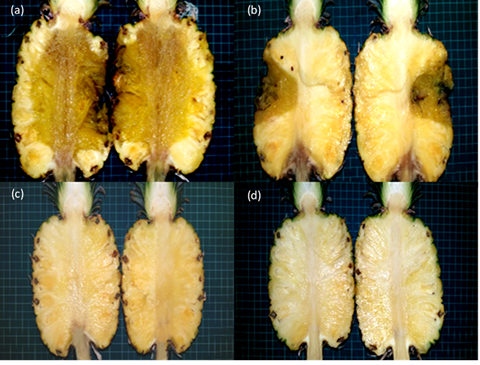
Fig. 1: Pathogenicity test of Thielaviopsis paradoxa on healthy MD2 pineapple fruits. (a) Typical black rot symptoms with soft, water-soaked lesions observed at wounded inoculation sites. (b) Localized rot symptoms developing at unwounded inoculation sites. (c) No visible symptoms on wounded control fruit inoculated with sterile PDA disc. (d) No symptoms on unwounded control fruit, confirming the absence of contamination or natural infection
A total of 103 fungal isolates were obtained from healthy pineapple tissues placed on PDA. These isolates were grouped based on similar culture characteristics as Trichoderma, Fusarium, Penicillium and Aspergillus. All fungal isolates were further screened as biological control agents against black rot pathogen in vitro. Results from the dual culture test indicated that an isolate morphologically identified as Trichoderma species gave the highest PIRG value of 98% compared to other microbial isolates after 7 days of incubation. The interaction between Trichoderma species and fungal pathogen was characterized by overgrowth of the antagonist on the pathogen's colony, followed by visible signs of pathogen suppression such as reduced mycelial density and discoloration at the interaction zone.
Characteristics of fungal pathogen and beneficial fungus
The fungal pathogen obtained from the pathogenicity test displayed morphological characteristics consistent with Thielaviopsis species when grown on PDA media. During the early growth stage, the mycelium appeared white and downy, gradually transitioning to a greenish- grey color upon sporulation (Fig. 2a). As the culture matured, the mycelium turned into a distinctive black velvet-like appearance (Fig. 2b). The culture also produced strong fruity odors that could be described as ethyl alcohol and ethyl acetate. Meanwhile, for microscopic characterization, abundant conidia and chlamydospores were observed, as shown in Fig. 2c, d and e. The conidia were cylindrical or barrel-shaped, forming long chains, that ranged from colorless to pale brown in appearance with sizes measuring 3.09 – 20.17 × 3.10–5.57 μm (n = 20). In addition, chlamydospores with pyriform (pear-shaped) and thick-walled brown color were observed, occurring either singly or in chains, with sizes ranging from 8.02 to 21.32 × 4.20 to 9.76 μm (n = 20). Furthermore, molecular identification using BLAST analysis confirmed that the sequence of the pathogen isolate showed 100% sequence homology to the Thielaviopsis paradoxa isolate PCC.02 with the GenBank accession no. HQ248205.1. Meanwhile, for BCA, the fungus was characterized as Trichoderma species with initial whitish mycelium that gradually turned dark green at maturity (Fig. 3a). Conidia, conidiophores and phialides were observed and the characteristics of these structures were matched with the description of Trichoderma by Siddiquee et al. (2017). The conidia were olive green in color and sub-globose in shape, the conidiophores exhibited verticillate branching with lageniform phialides positioned terminally (Fig. 3b, c). Meanwhile, chlamydospores were found both intercalary and terminally (Fig. 3d). The species of Trichoderma was then confirmed using ITS rRNA gene phylogenetic analysis and BLAST results showed the BCA sequence was 99-100% closely related to the sequence of Trichoderma asperellum strain ANP (accession number JX913783.1).
Effectiveness of T. asperellum against T. Paradoxa in vivo
Table 3: Disease severity index of pineapple fruits (var. MD2) that were inoculated with Thielaviopsis paradoxa and treated with Trichoderma asperellum suspension
|
Observation Days |
Disease Severity Index (%) |
||
|
T1 (Positive Control); Mean ± SD |
T4 (Curative Treatment); Mean ± SD |
T5 (Preventive Treatment); Mean ± SD |
|
|
0 |
0.000 |
0.000 |
0.000 |
|
2 |
67 ± 0.57 |
33 ± 0.75 |
33 ± 0.830 |
|
4 |
83 ± 0.29 |
44 ± 0.62 |
33 ± 0.580 |
|
6 |
83 ± 0.39 |
83 ± 0.80 |
61 ± 0.96 |
|
8 |
100 ± 0.00 |
100.00 ± 0.00 |
670 ± 0.58 |
|
10 |
100 ± 0.00 |
100.00 ± 0.00 |
670 ± 0.82 |
Note: T2 and T3 were excluded from Table 3 because their mean values were zero, indicating no disease symptoms. Mean ± standard deviation
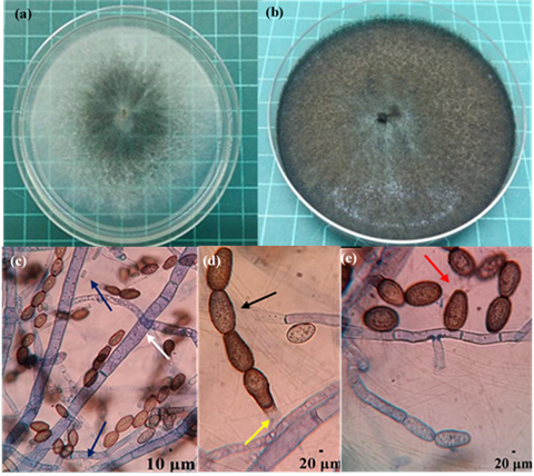
Fig. 2: Morphological and microscopic characteristics of Thielaviopsis paradoxa, causal pathogen of black rot in pineapple. (a) Four-day-old colony on PDA showing white, downy mycelium transitioning to greenish gray. (b) Ten-day-old colony displaying a mature black, velvet-like appearance typical of T. Paradoxa. (c) Conidiogenous cells and phialoconidia (blue arrows) along with septate hyphae (white arrow) observed under light microscope (400x magnification). (d) Chains of aleuroconidia (black arrow) and conidiophores (yellow arrow). (e) Singly occurring chlamydospores with thick walls and pyriform shape (red arrow)
The in vivo findings supported the in vitro results, confirming the effectiveness of T. asperellum in suppressing black rot disease in pineapple fruits. The Disease Severity Index (DSI) values for Treatments T1, T4 and T5 are summarized in Table 3. On day 0, no disease symptoms were observed across all treatments (DSI = 0.00; Scale 0) (Fig. 4a). Disease symptoms began to develop from peduncles to fruit flesh in treatments T1, T4, and T5 by day 2 (Fig. 4b). A significant difference in DSI was recorded between T1 and T4, as well as T1 and T5 (P < 0.05). T1 exhibited a DSI of 67% (Scale 6), whereas both T4 and T5 recorded 33% (Scale 5). Control treatments T2 and T3 remained symptomless. By day 4, the DSI in T1 increased to 83.3% (Scale 6), indicating advanced disease development (Fig. 4c). On the other hand, T4 and T5 showed slower disease progression, with DSI values of 44% and 33%, respectively. On day 6, disease severity in T4 escalated to 83% (Scale 6), matching that of T1 (Fig. 4d). In contrast, T5 showed a significantly lower DSI of 61% (P<0.05 vs. T1). This finding indicates that preventive treatment, in which the application of T. asperellum had occurred prior to the pathogen was able to slow down the disease infection compared to curative treatment, in which T. asperellum was applied after the pathogen. Similar trends were observed on days 8 and 10, DSI in T1 and T4 reached 100% (Scale 6) (Fig. 4e and f). Meanwhile, T5 showed a moderate increase from 61% to 67% on day 8 and remained constant through day 10. Throughout the study, T2 and T3 showed no visible symptoms and consistently recorded a DSI of 0% (Scale 0).
Table 4: AUDPC and the percentage of disease reduction (DR) for the studied treatments
|
Treatment |
AUDPC Value |
DR (%) |
|
T1 (T. paradoxa alone) |
7.66 |
0 |
|
T2 (SDW only) |
0 |
100 |
|
T3 (T. asperellum alone) |
0 |
100 |
|
T4 (Curative) |
6.22 |
18.83 |
|
T5 (Preventive) |
4.56 |
40.57 |
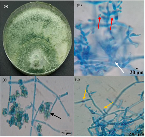
Fig. 3: Morphological and microscopic characteristics of Trichoderma asperellum, the selected BCA. (a) Seven-day-old colony on PDA exhibiting initial whitish mycelium then turned dark green upon sporulation. (b) Verticillate conidiophores (white arrows) with terminal lageniform phialides (red arrows). (c) Subglobose olive-green conidia (black arrow). (d) Chlamydospores formed both intercalary and terminally along the hyphae (yellow arrows), visualized under 400x magnification
Disease progression analysis
The severity of black rot disease in MD2 pineapple fruits was further evaluated using the Area Under the Disease Progress Curve (AUDPC). The effectiveness of T. asperellum spore suspension was quantified by the percentage of disease reduction (%DR), derived from the AUDPC values. As presented in Table 4, treatment T5 recorded the lowest AUDPC value (4.56 units²), indicating the highest efficacy in suppressing disease progression, with a 41% reduction in disease severity. In contrast, T1 exhibited the highest AUDPC value (7.66 units²), reflecting the absence of disease suppression. Treatment T4 showed a moderate AUDPC of 6.22 units², corresponding to an 19% reduction in disease severity, suggesting limited effectiveness once the infection was established. Both negative controls, T2 and T3 recorded no symptoms (AUDPC = 0.00), confirming the absence of infection and validating the experimental setup.
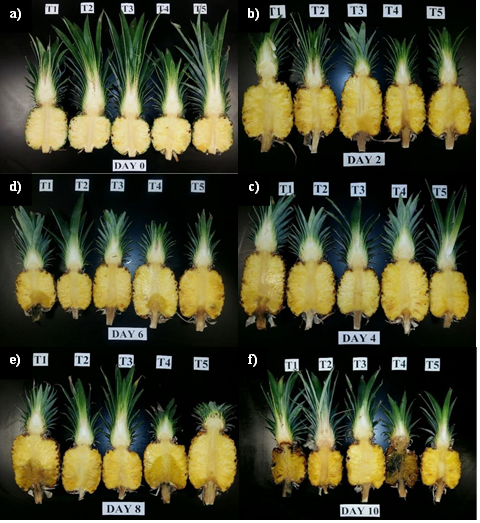
Fig. 4: Disease progression of black rot in MD2 pineapple over a 10-day period following treatment with Trichoderma asperellum and Thielaviopsis paradoxa. (a) No visible symptoms on Day-0 in all treatments. (b) Early symptoms of water-soaked lesions observed in pathogen-inoculated treatments on Day-2. (c) Disease progression evident in T1 and T4 on Day-4. (d-e) Severe lesion development in T1, T4 and comparatively limited progression in T5 on Day-6 and 8. (f) highly severe in T1 and T4 on Day-10. T2 and T3 consistently showed no disease symptoms
Discussion
The present study demonstrated that T. asperellum has potential as a biological control agent against T. paradoxa, the causal agent of black rot in pineapple var. MD2. In vitro screening using dual culture assays revealed that T. asperellum significantly inhibited the radial growth of T. paradoxa, with a high PIRG of 98%. This discovery aligns with previous studies indicating that T. asperellum exhibited antagonistic activity via mechanisms including mycoparasitism, nutrient competition, and the synthesis of antifungal metabolites (Selva et al. 2024). In vivo experiments also confirmed the efficacy of T. asperellum in reducing black rot disease symptoms in pineapple var. MD2 Treatments using T. asperellum clearly slowed down the course of the disease, particularly in the preventive application (T5), which regularly showed lower disease severity index (DSI) values over the 10-day incubation period. Whereas 100% was found in the positive control (T1), fruits treated with T. asperellum before pathogen inoculation (T5) showed a maximum DSI of 67%. These findings suggest that early application of T. asperellum can significantly mitigate the progression of black rot in MD2 pineapple fruits and aligned with previous studies that emphasize the importance of timing applications, showing that pre-inoculation treatments with biocontrol agents significantly reduce disease severity in crops such as bananas (Rahman et al. 2021), strawberries (Liu et al. 2024), and potatoes (Metz and Hausladen 2022). Conversely, the curative treatment (T4), in which T. asperellum is applied following pathogen inoculation, showed only partial control of disease progression, with less than a 20% decrease. This finding suggests that biocontrol action post-infection of T. asperellum loses effectiveness once the pathogen has colonized host tissues. Wijesinghe et al. (2010) also noted this phenomenon and emphasized the need for early biocontrol agent applications to maximize their efficacy against fungal pathogens. The rapid colonization capacity of T. paradoxa, which starts within 60 min post-inoculation, can be attributed to the reduced efficacy of the therapeutic intervention since it exceeds the colonization of the antagonist administered later on the onset of an infection. The preventive mechanism observed in treatment T5 underscores the protective attributes of T. asperellum, endorsing its application as a prophylactic measure in postharvest management. These results coincide with studies by Janisiewicz and Korsten (2002), which show that, to control wound-invading pathogens, postharvest treatments of antagonists are most effective if administered either beforehand or immediately following harvesting. The mechanism of T. asperellum entails the synthesis of antifungal agents, including 6-pentyl-pyrone (6-PP), which is recognized for its antifungal efficacy (Hamrouni et al. 2021). The pronounced suppressive impacts noted in the present study may also be ascribed to these secondary metabolites, alongside direct antagonism and competitive exclusion. Numerous research studies have indicated that T. asperellum can rapidly colonize sites of wounds, which prevents the development of pathogens (Ladjouzi et al. 2023). These findings from studies are especially important in the context of reducing dependence on chemical fungicides, which are now the mainstay for controlling black rot in pineapples, despite having environmental and human health issues. Nevertheless, to maximize the efficacy of T. asperellum and guarantee consistent results when using this Trichoderma-based product in postharvest disease management strategies for pineapples, additional research on the formulation and delivery of this BCA to plant systems is crucially required.
Conclusion
Trichoderma asperellum was identified as a useful biological control agent against T. paradoxa, the pathogen causing black rot disease in pineapple fruits. The ability of T. asperellum to reduce black rot disease in pineapple var. MD2 was validated by both in vivo and vitro experiments, which demonstrated a significant reduction in disease progression and strong antagonistic activity against the pathogen. Administering T. asperellum before pathogen inoculation revealed low disease severity and AUDPC values, thereby confirming its possible pre-infection protection ability. As a conclusion, these research findings provide a promising foundation for developing environmentally friendly alternatives to chemical fungicides in postharvest disease management of pineapple, particularly for export-quality Malaysian MD2 fruits. Further research into formulation development and field-scale applications is recommended to enhance the practical use of T. asperellum as a biocontrol agent.
Acknowledgements
The authors acknowledge the financial support provided by the Ministry of Higher Education (MOHE), Malaysia, through the Fundamental Research Grant Scheme (FRGS) (File no. FRGS/1/2016/WAB01/UITM/02/4).
Authors Contribution
NFMH conceived the study, designed and performed experiments, analyzed the data and drafted the manuscript. ZS provided the research idea, co-designed the experiments, and critically revised the manuscript. ACA contributed to data analysis, participated in the interpretation of the results, and assisted in manuscript refinement.
Conflict of Interest
All authors declare no conflict of interest.
Data Availability
Data presented in this study will be available on a fair request to the corresponding author.
Ethics Approval
Not applicable to this paper.
References
Ali A, A Javaid, A Shoaib, IH Khan (2020). Effect of soil amendment with Chenopodium album dry biomass and two Trichoderma species on growth of chickpea var. Noor 2009 in Sclerotium rolfsii contaminated soil. Egypt J Biol Pest Contr 30:1-9
Ali HZ, K Nadarajah (2013). Evaluating the efficacy of Trichoderma isolates and Bacillus subtilis as biological control agents against Rhizoctonia solani. Res J Appl Sci 8:72–81
Anbalagan SA, S Appusamy, PV Kumaresan, G Chellappan, S Narayanan, A Rangasamy, K Perveen, NA Bukhari, R Sayyed (2025). Deciphering the biocontrol potential of Trichoderma asperellum (Tv1) against Fusarium‐Nematode wilt complex in tomato. J Basic Microbiol 65:2400595
Baiswar P, TR Borah, AR Singh (2021). Postharvest diseases of pineapple and their management. In: Postharvest handling and diseases of horticultural produce, pp:287–294. Singh D, RR Sharma, V Devappa, D Kamil (Eds.). CRC Press, Boca Raton, USA
Cai F, K Dou, P Wang, K Chenthamara, J Chen, IS Druzhinina (2022). The current state of Trichoderma taxonomy and species identification. In: Advances in Trichoderma biology for agricultural applications, pp:3–35. Amaresan N, A Sankaranarayanan, MK Dwivedi, IS Druzhinina (Eds.). Springer International Publishing, Cham, Switzerland
Camacho-Luna V, AM Pizar-Quiroz, AA Rodríguez-Hernández, M Rodríguez-Monroy, G Sepúlveda-Jiménez (2023). Trichoderma longibrachiatum, a biological control agent of Sclerotium cepivorum on onion plants under salt stress. Biol Contr 180:105168
FAO (2024). Major Tropical Fruits Market Review. Preliminary results 2024. Food and Agriculture Organization of the United Nations. Available at: https://openknowledge.fao.org/handle/20.500.14283/cd3818en (Accessed: March 20, 2024)
Granada D, L Lopez-Lujan, S Ramirez-Restrepo, J Morales, C Pelaez-Jaramillo, G Andrade, JC Bedoya-Pérez (2020). Bacterial extracts and bioformulates as a promising control of fruit body rot and root rot in avocado cv. Hass. J Integr Agric 19:748–758
Gutiérrez-Chávez A, L Robles-Hernández, BI Guerrero, AC González-Franco, G Medina-Pérez, AA Acevedo-Barrera, J Hernández-Huerta (2025). Potential of Trichoderma asperellum as a growth promoter in hydroponic lettuce cultivated in a floating-root system. Plants 14:1-18
Hamrouni R, N Dupuy, J Molinet, S Roussos (2021). Trichoderma species: novel metabolites active for industry and biocontrol of mycotoxigenic fungi. In: Mycotoxins in Food and Beverages, pp:1–25. Montet D, C Brabet, S Schorr-Galindo, RC Ray (Eds.). CRC Press, Boca Raton, USA
Hubert J, M Nesvorná, J Kopecký (2014). The effect of Tyrophagus putrescentiae on Fusarium poae transmission and fungal community in stored barley in a laboratory experiment. Ins Sci 21:65–73
Janisiewicz WJ, L Korsten (2002). Biological control of postharvest diseases of fruits. Annu Rev Phytopathol 40:411–441
Javaid A, L Afzal, A Shoaib (2017). Biological control of charcoal rot of mungbean by Trichoderma harzianum and shoot dry biomass of Sisymbrium irio. Plant Danin 35:1-12
Khan BA, MA Nadeem, H Nawaz, MM Amin, GH Abbasi, M Nadeem, M Ali, M Ameen, MM Javaid, R Maqbool, M Ikram (2023). Pesticides: impacts on agriculture productivity, environment, and management strategies. In: Emerging contaminants and plants: Interactions, adaptations and remediation technologies, pp:109–134. Aftab T (Ed.). Springer International Publishing, Cham, Switzerland
Khan IH, A Javaid (2020). In vitro biocontrol potential of Trichoderma pseudokoningii against Macrophomina phaseolina. Intl J Agric Biol 24:730–736
Khan IH, A Javaid, D Ahmed (2021). Trichoderma viride controls Macrophomina phaseolina through its DNA disintegration and production of antifungal compounds. Intl J Agric Biol 25:888–894
Kuruppu M, Y Siddiqui, LL Kong, K Ahmed, A Ali (2021). First report of postharvest stem end rot disease on MD2 pineapple fruits caused by Neoscytalidium dimidiatum in Malaysia. Plant Dis 105:1-1
Ladjouzi R, M Bachir-Bey, F Zaidi, R Yahiaoui-Zaidi (2023). Biological control of soft rot induced by Dickeya solani with Trichoderma asperellum on potato tubers: Relationship with susceptibility of variety. Acta Phytopathol Entomol Hung 58:168–179
Liu P, R Yang, Z Wang, Y Ma, W Ren, D Wei, W Ye (2024). Biocontrol potential of Trichoderma asperellum CMT10 against strawberry root disease. Horticulturae 10:1-16
Masood A, S Saeed, N Iqbal, MT Malik, MR Kazmi (2010). Methodology for the evaluation of symptoms severity of mango sudden death syndrome in Pakistan. Pak J Bot 42:1289–1299
Metz N, H Hausladen (2022). Trichoderma spp. as potential biological control agent against Alternaria solani in potato. Biol Contr 166: 104820
Rahman SSMSA, NAIM Zainudin, NAA Aziz (2021). Evaluation of Trichoderma asperellum B1902 in controlling Fusarium wilt of cavendish banana cultivar. Sains Malays 50:2549–2561
Rebitanim NA, MM Hanafi, AS Idris, SNA Abdullah, H Mohidin, NZ Rebitanim (2020). GanoCare® improves oil palm growth and resistance against Ganoderma basal stem rot disease in nursery and field trials. Biomed Res Intl 2020:1–16
Rivera-Mendez W, M Obregon, ME Moran-Diez, R Hermosa, E Monte (2020). Trichoderma asperellum biocontrol activity and induction of systemic defenses against Sclerotium cepivorum in onion plants under tropical climate conditions. Biol Contr 141:104145
Rohrbach KG, MW Johnson (2003). Pests, diseases and weeds. In: The pineapple: botany, production and uses, pp:203–251. Bartholomew DP, RE Paull, G Rohrbach (Eds.). CABI publishing, Wallingford, UK
Sapak Z, ANMF Mahadeven, MHN Farhana, S Norsahira, AWM Zafri (2021). A review of common diseases of pineapple: The causal pathogens, disease symptoms, and available control measures. Food Res 5:1–14
Seekham N, N Kaewsalong, A Jantasorn, T Dethoup (2024). Biological control of Corynespora leaf fall disease in rubber by endophytic Trichoderma spp. under field conditions. Eur J Plant Pathol 170:207–218
Selva AA, VK Parthiban, A Sudha, C Gopalakrishnan, N Swarnakumari, R Anandham (2024). Antifungal and plant-growth promoting potency of Trichoderma asperellum against Fusarium wilt on tomato. J Plant Pathol 2024:1–13
Shoaib A, M Munir, A Javaid, AZ Awan, M Rafiq (2018). Anti-mycotic potential of Trichoderma spp. and leaf biomass of Azadirachta indica against the charcoal rot pathogen Macrophomina phaseolina (Tassi) Goid in cowpea. Egypt J Biol Pest Contr 28:1-7
Siddiquee S, SN Shafawati, L Naher (2017). Effective composting of empty fruit bunches using potential Trichoderma strains. Biotechnol Rep 13:1–7
Skidmore AM, CH Dickinson (1976). Colony interactions and hyphal interference between Septoria nodorum and phylloplane fungi. Trans Brit Mycol Soc 66:57–64
Sundram S, F Abdullah, ZAM Ahmad, UK Yusuf (2008). Efficacy of single and mixed treatments of Trichoderma harzianum as biocontrol agents of Ganoderma basal stem rot in oil palm. J Oil Palm Res 20:470–483
Wijesinghe CJ, RW Wijeratnam, JKRR Samarasekara, RLC Wijesundera (2011). Development of a formulation of Trichoderma asperellum to control black rot disease on pineapple caused by (Thielaviopsis paradoxa). Crop Prot 30:300–306
Wijesinghe CJ, RW Wijeratnam, JKRR Samarasekara, RLC Wijesundera (2010). Biological control of Thielaviopsis paradoxa on pineapple by an isolate of Trichoderma asperellum. Biol Contr 53:285–290
Online : 1814-9596
Print : 1560-8530