


Indexing


Journal Cover

Supranutritional Selenium Supplementation Modulated Lipopolysaccharide Provoked Inflammatory Injury in Colon of Goat Fed Grain Rich Diet
Abstract
The current research was designed to examine the role of extra selenium (Se) supplemented against high grain diet induced stress mediated inflammatory response leading to epithelial injury in the colon of goat. Eighteen goats were divided into three dietary groups: one received a low-grain diet (LG; grain to forage ratio of 35:65), another was fed a high-grain diet (HG; grain to forage ratio of 65:35), and the third group was provided a high-grain diet supplemented with selenium (HG-Se; 65:35 plus 0.5 mg Se per kg of diet). At the end of tenth week experiment period, compared with low grain, high grain diet elevated (P < 0.05) colonic and plasma lipopolysaccharide (LPS) concentration. Colonic epithelial injury accompanied with enhanced (P < 0.05) melondialdehyde (MDA) and diminishing (P < 0.05) the activities of total antioxidant status (T-AOS) was observed in high grain diet than low grain diet. Moreover, the HG diet led to a significant increase (P < 0.05) in the levels of plasma acute phase proteins (APPs), including serum amyloid A (SAA), lipopolysaccharide-binding protein (LBP) and haptoglobin (Hp), accompanied by changes in the colonic mRNA expression of immune-related genes such as toll-like receptor 4 (TLR-4), cluster of differentiation 14 (CD-14), tumor necrosis factor-alpha (TNF-α), nuclear factor kappa B (NF-κB), and interleukins IL-1, IL-6, IL-10, and IL-13. Conversely, surplus Se supplementation lessens LPS level in colon and blood thus alleviated colonic epithelial injury in high grain-Se diet. Besides, Se supplemented high grain diet modulated plasma APPs and inflammatory genes in goat colon. In conclusion, high grain diet-induced colonic inflammation was associated with TLR-4- MYD88 signaling pathway. Conversely, extra Se supplementation alleviated high grain diet induced stress mediated inflammatory response through TLR-4- MYD88 signaling pathway thus reduced colonic injury in the goats.
Keywords: High grain diet; Selenium; Lipopolysaccharide; Acute phase response; Inflammation
Supranutritional Selenium Supplementation Modulated Lipopolysaccharide Provoked Inflammatory Injury in Colon of Goat Fed Grain Rich Diet
Nabi Bux Solangi1, Saba Parveen Samo1, Moolchand Malhi1*, Jamila Soomro1, Fayaz Ali Ujjan2 and Muhammad Awais Soomro3
1Department Veterinary Physiology and Biochemistry, Sindh Agricultural University, 70060 Tando Jam, Pakistan
2Vaccine production unit, 70060 Sindh Tandojam, Pakistan
3Department of Veterinary Physiology and Biochemistry, Shaheed Benazir Bhutto University of Veterinary and Animal Science 67210 Sakrand, Pakistan
*For correspondence: mcmalhi@sau.edu.pk; Mobile: +92-3361241635
Received 27 June 2025; Accepted 02 August 2025; Published online 22 September 2025
Editor: Zafar Iqbal
Abstract
The current research was designed to examine the role of extra selenium (Se) supplemented against high grain diet induced stress mediated inflammatory response leading to epithelial injury in the colon of goat. Eighteen goats were divided into three dietary groups: one received a low-grain diet (LG; grain to forage ratio of 35:65), another was fed a high-grain diet (HG; grain to forage ratio of 65:35), and the third group was provided a high-grain diet supplemented with selenium (HG-Se; 65:35 plus 0.5 mg Se per kg of diet). At the end of tenth week experiment period, compared with low grain, high grain diet elevated (P < 0.05) colonic and plasma lipopolysaccharide (LPS) concentration. Colonic epithelial injury accompanied with enhanced (P < 0.05) melondialdehyde (MDA) and diminishing (P < 0.05) the activities of total antioxidant status (T-AOS) was observed in high grain diet than low grain diet. Moreover, the HG diet led to a significant increase (P < 0.05) in the levels of plasma acute phase proteins (APPs), including serum amyloid A (SAA), lipopolysaccharide-binding protein (LBP) and haptoglobin (Hp), accompanied by changes in the colonic mRNA expression of immune-related genes such as toll-like receptor 4 (TLR-4), cluster of differentiation 14 (CD-14), tumor necrosis factor-alpha (TNF-α), nuclear factor kappa B (NF-κB), and interleukins IL-1, IL-6, IL-10, and IL-13. Conversely, surplus Se supplementation lessens LPS level in colon and blood thus alleviated colonic epithelial injury in high grain-Se diet. Besides, Se supplemented high grain diet modulated plasma APPs and inflammatory genes in goat colon. In conclusion, high grain diet-induced colonic inflammation was associated with TLR-4- MYD88 signaling pathway. Conversely, extra Se supplementation alleviated high grain diet induced stress mediated inflammatory response through TLR-4- MYD88 signaling pathway thus reduced colonic injury in the goats.
Keywords: High grain diet; Selenium; Lipopolysaccharide; Acute phase response; Inflammation
Introduction
Grain-based diet usually fed to the ruminants is rich in easily fermentable carbohydrates, high in starch and energy. Long term feeding of grain rich diet results in the development of acidic medium in the gastrointestinal tract (GIT) that provokes acid intolerant gram negative bacteria to release large number of endotoxins such as lipopolysaccharides (LPS) (Chen et al. 2023). Concurrently, luminal accumulation of LPS compromises GIT barrier resulting in release of LPS to the blood through impaired ruminal epithelium (Tao et al. 2015) and hindgut (Samo et al. 2020) resulting the development of inflammatory response in various tissue and organs of the body. Though, the LPS translocation occurs from rumen as well as hindgut (Colon and cecum) but being mono-layered in structure and deficient in natural buffering capacity, hindgut is easily predisposed to abnormal conditions in the lumen, hence considered as abundant site for the translocation of LPS (Tao et al. 2017). Further studies suggested that accumulation of LPS due to grain rich diet causes jejunal and ileal epithelial injury in dairy cows (Lai et al. 2022) increases pro-inflammatory gene expression in the rumen (Liu et al. 2013) and hindgut (Tao et al. 2014), modulates acute phase protein (APPs) concentration in blood (Chang et al. 2015) hence leading to the development of local and systemic inflammation.
Lipopolysaccharide (LPS) as a potent inflammatory mediator, triggers and binds with toll like receptors (TLR4) on the membrane of host intestinal epithelial cells and triggers myeloid differentiating factor 88 (MyD88). Pro-inflammatory cytokines are being stimulated by the TLR4/MyD88 pathway by mediating the activation of NF-κB to regulate stimulation of IL-1β, IL-6 and TNF-α (Kawai and Akira 2009). Additionally, in dairy goats fed a grain-rich diet, LPS induced stress resulted in elevated levels of acute phase proteins (APPs), including serum amyloid A (SAA) (Jia et al. 2014), as well as increased concentrations of haptoglobin (Hp) and lipopolysaccharide-binding protein (LBP) (Chishti et al. 2020) in dairy goats fed grain rich diet. The plasma levels of LBP significantly increases up to 200% in grain fed goats that facilitate LPS binding to the receptors of cell membrane, causes the release of other APPs into the blood hence triggers the series of events towards local and systemic inflammatory response (Chang et al. 2015).
Selenium (Se) documented as vital micro- nutrient in the mammalian diet due to its various biochemical properties (Sherlock et al. 2020; He et al. 2022). However, the affects primarily hinge on its biological availability and chemical form (Zeng 2009). At nutritional level (0.1-0.5 mg/kg diet), exhibits broad physiological functions comprising defensive mechanism against oxidative damages, prevention of cell death and modulation of immune system (Hu et al. 2018). Furthermore, extra Se (10 - 20 folds higher than recommended) retained supplementary Se thus pertained higher antioxidant capability in animal tissues with less adverse effects on wellbeing of animals (Razo-Rodriguez et al. 2013). Particularly, addition of surplus amount of Se in the diet during stressed conditions exerted the protective effect. The additional level of Se (0.6 - 0.7 mg/kg diet) showed anti-apoptotic and anti-inflammatory effects in goats during stressed condition, attenuating grain rich diet induced oxidative injuries in colon (Samo et al. 2020) and liver (Shah et al. 2022). Besides, Se reduces stress-mediated natural defenses through diminution of APPs in cattle during pre and postpartum (Gong and Xiao 2021). Reports also suggested that in sheep and pigs, supranutritional levels of Se supplementation during stress condition improved anti-oxidant status and alleviated inflammation (Chen et al. 2020; Liu et al. 2021). Earlier researches have revealed that protective effect of Se against heat stress (He et al. 2022) and LPS- induced oxidative stress (OS) and inflammation in pigs, mice and broilers is through activation of TLR4 inflammatory pathway (Qu et al. 2020).
Since, insufficiency of Se prevents the growth and functions of immune organs triggering inflammatory diseases (Stranges et al. 2011). The Se deficiency leading to lowered glutathione peroxidase (GSH-Px) activity, thus stimulated LPS resulting in oxidative injuries caused by inflammation in sheep, goat and mice (Sherlock et al. 2020). Numerous findings have shown that Se enhance immune status and exert anti-inflammatory action, also inhibits LPS induced expressions of pro-inflammatory genes in primary epithelial cells in mice (Zhang et al. 2015), showing significance of Se in controlling inflammation due to increase of LPS concentration thus specifies role of Se during stressed environment. Thus, the findings lead to hypothesize that supplementing surplus amount of Se would have protecting effects over colonic damages induced by grain rich diet by increasing antioxidant properties via hindering inflammatory pathway in colon. Hence, existing study estimated the role of extra dose of Se supplementation against grain rich diet-induced OS leading to inflammatory status in the colon of goats.
Material and Methods
Experimental design
The current research was permitted by Ethical Committee of Sindh Agriculture University Tandojam, Pakistan (No. DAS/1956/ of 2024 Dated: 12.08.2024 in 160th meeting of BASAR held on 06.08.2024, under resolution no. 160th BASAR-09). Trail was performed on eighteen female goats allocated into three groups after four weeks for acclimatization; each group contained six goats, penned into ten Sq. ft. i.e., 2.5×). The goats were fed low grain (LG, i.e., grain forage ratio 35 : 65), whereas high grain (HG ratio 65:35) and HG supplemented with Se (HG-Se) diets, twice a day (morning and evening) with free access of water. The Se concentrations in the diets were determined using inductively coupled plasma optical emission spectrometry (ICP-OES Optima 2100-DV, Perkin Elmer) (Taylor et al. 2010). The Se concentration analyzed was 0.035 mg Se kg-1 diet, 0.15 mg Se kg-1 diet in low grain and high grain respectively, Additional amount of 0.115 mg Se kg-1 diet in low grain and 0.5 mg Se kg-1 diet in hig grain-Se group was added to accomplish 0.15 mg Se kg-1 diet in low grain and high grain groups where as 0.65 mg Se kg-1 diet in high grain-Se. Added organic Se used in the form of selenium yeast (SY) according to our previously published research (Samo et al. 2020). Composition of diets given to animals is illustrated in Table 1.
Slaughtering, organ isolation and sample collection
At the end of the 10-week experimental trial, 10 mL blood samples collected from jugular vein in K3EDTA and plan vacutainer tubes. Samples were centrifuged at 5,000 × g for 20 min at 4°C within 20 min of collection. The resulting plasma was stored at −20°C for later analysis. Thereafter, all animals were sacrificed, GIT was exteriorized immediately through midline incision and hindgut was isolated at ileocecal junction. Colon was carefully isolated from hindgut, collected into clean tub. The digesta after removal and collected in a container for LPS analysis. Histological samples were collected and fixed using 10% formaldehyde for analysis. The epithelial layer was then carefully separated from the opened wall using a glass slide, transferred into an Eppendorf tube, and stored frozen until gene expression analysis.
Table 1: Formulated dietary treatments fed to experimental animals
|
Items |
Treatments |
|
|
LG |
HG |
|
|
Ingredients (% of DM) |
||
|
Corn |
25.6 |
25 |
|
Wheat bran |
- |
30.7 |
|
Soybean meal |
7.4 |
2.2 |
|
Rapeseed meal |
- |
4 |
|
Lime stone |
0.5 |
1.5 |
|
DCP |
0.8 |
0.7 |
|
Salt |
0.4 |
0.4 |
|
Mineral Premix1 |
0.4 |
0.4 |
|
Nutrients (% DM) |
||
|
Energy (MJ/kg) |
5.96 |
6.32 |
|
EE |
2.89 |
3.66 |
|
NDF |
38.11 |
35.36 |
|
ADF |
25.26 |
18.33 |
|
NFC |
46.06 |
37.39 |
DCP = Digestible crude protein, EE = Ether extract, NDF = Nutrient detergent fiber, ADF = acid detergent fiber, NFC = Nitrogen free extract. 1Per kg of premix = Vitamin A 6000 U; Vitamin D2 500 U; Vitamin E 80 mg; Cu 6.25 mg; Fe 62.5 mg; Zn 62.5 mg; Mn 50 mg; I 0.125 mg; Co 0.125 mg; Mo 0.125 mg
Table 2: Primers for real time RT-PCR
|
Gene |
Primer sequence 5’ to 3’ |
Accession number |
Size (bp) |
|
GAPDH |
GGGTCATCATCTCTGCACCT GGTCATAAGTCCCTCCACGA |
HM043737.1 |
180 |
|
IL-6 |
CCAATCTGGGTTCAATCAGG ACCCACTCGTTTGAGGACTG |
D86569.1 |
241 |
|
IL-10 |
TTAAGGGTTACCTGGGTTGC CCCTCTCTTGGAGCATATTGA |
DQ837159.1 |
239 |
|
IL-13 |
CAGTGTCATCCAAAGGACCAAG CGGACGTACTCACTGGAAACC |
NM174089 |
248 |
|
IL-1 |
GAAGAGCTGCACCCAACA CAGGTCATCATCACGGAAG |
D63351.1 |
172 |
|
TLR-4 |
GTTTCCACAAGAGCCGTAA TGTTCAGAAGGCGATAGAGT |
JQ342090.1 |
195 |
|
TNF-α |
CAAGTAACAAGCCGGTAGCCC CCTGAAGAGGACCTGCGAGTAG |
AF276985.1 |
173 |
|
NF-kB |
ACGATCGTCACCGGATTGAG GGTGCTGAGAGATGGCGTAA |
XM_005699996.1 |
194 |
|
MYD88 |
ACAAGCCAATGAAGAAAGAG GAGGCGAGTCCAGAACC |
JQ308783.1 |
98 |
|
CD14 |
CCGTTCAGTGTATGGTTGCC TGCTTCGGGTCGGTGTT |
NM_001077209.1 |
239 |
Equal volume of normal saline was carefully added to the colonic digesta, followed by vortexing and centrifugation at 3000 × g for 15 min, collected samples then analyzed for LPS concentration using the Chromogenic End-point Tachypleus Amebocyte Lysate Assay Kit (Chinese Horseshoe Crab Reagent Manufactory Co. Ltd., Xiamen, China).
Blood samples assay
Histo-morphological and histo-morphometric analysis
The colon tissue samples previously fixed in formalin and further processed for the evaluation of histo-morphological characteristics. Following dehydration, clearing, and paraffin embedding, tissue sections were cut into 4-µm slices using a microtome. The specimens were then stained using the standard hematoxylin and eosin (H&E) technique. For each goat, at least 10 replications were measured.
Analysis of T-AOS and MDA concentration
The epithelial tissue samples were homogenized in standard PBS solution. Total oxidant status (T-AOS) and MDA concentration in colonic epithelium were analyzed by using ELISA kits. The manufacturer’s instructions were strictly followed while performing the procedures.
Total RNA isolation and gene expression
Statistical analysis
Statistical analysis was conducted using SPSS version 16.0 (Stata Soft, Tulsa, OK, USA). Data are expressed as mean values ± standard error, and differences were considered statistically significant at P < 0.05.
Table 3: The effect of LG, HG and HG-Se diets on LPS concentration in rumen, colon and plasma of goat
|
Items |
Diets |
||
|
LG |
HG |
HG-Se |
|
|
LPS (EU/ml) |
|
|
|
|
Colon |
21023 ± 949.41c |
35279 ± 287.7a |
25622 ± 945.47b |
|
Plasma |
0.0875 ± 0.998c |
0.9375 ± 0.0457a |
0.328 ± 0.0658b |
LPS = lipopolysaccharide. Values are mean ± S.E and a, b, c values with different superscripts were considered significant at P < 0.05
Table 4: The effect of LG, HG and HG-Se diets on acute phase proteins in plasma of goat
|
Parameters |
Diets |
||
|
LG |
HG |
HG-Se |
|
|
SAA (µg/ml) |
10.532 ± 1.511c |
81.412 ± 1.02a |
27.527 ± 1.79b |
|
Hp (µg/ml) |
235.00 ± 14.629b |
377.50 ± 37.881a |
351.00 ± 16.14a |
|
LBP (µg/ml) |
17.375 ± 1.25b |
67.475 ± 1.98a |
24.800 ± 4.94b |
SAA = serum amyloid A, Hp = hepatoglobin, LBP = lipopolysacchride binding protein. Values are mean ± S.E and a, b, c values with different superscripts were considered significant at P < 0.05
Results
LPS concentration
The tissue and plasma LPS concentration significantly amplified (P < 0.05) for goats fed the HG diet compared to those on the LG diet (Table 3). Nevertheless, addition of Se in high grain diet showed significant decrease (P < 0.05) in LPS concentration in tissue, plasma by 27.37% and 65.01% respectively in high grain-Se as compared to high grain diet.
Histomorphology of colonic epithelium
The epithelium of the goats fed high grain diet exhibited severe damages to the colonic mucosa described by crypt necrosis and inflammatory cell infiltration (Fig. 1B) as compared to normal epithelium showed by low grain diet (Fig. 1A). Whereas, addition of Se improved high grain diet induced damaging changes and exhibited slight mucosal damages in high grain-Se diet (Fig. 1C) as compared to high grain diet.
Plasma acute phase proteins
The concentrations of all measured plasma APPs, i.e., SAA, Hp, and LBP, were significantly higher (P < 0.05) in goats fed the high grain diet compared to those on the low grain diet (Table 4). Conversely, supplementation of Se with high grain diet diminished significantly (P < 0.05) the concentration of SAA by 66.18% and LBP by 63.24% in high grain-Se as compared to high grain diet. The values of Hp were non-significant (P > 0.05) between high grain and high grain Se diets.
T-AOS and MDA concentration
Total antioxidant status (T-AOS) as shown in Fig. 1 (A) significantly decreased (P < 0.05) by 75% with concurrent increase in melondialdehyde (MDA) concentration (Fig. 1B) by 47.7% in high grain as compared to low grain group. Addition of Se to high grain diet showed significant increase (P < 0.05) in T-AOS by 400% and decrease in MDA concentration by 27.90% in high grain-Se groups as compared to high grain. However, the values of T-AOS were non-significant (P > 0.05) between low grain and high grain-Se diets.
Expression of inflammatory genes in colonic epithelium
The mRNA expression levels of the pro-inflammatory cytokines interleukin 1 (IL-1) and interleukin 6 (IL-6) were increased significantly (P < 0.05) as shown in (Fig. 2A) by 0.9-folds and 1.1-folds and expression level of anti-inflammatory IL-10 and IL-13 decreased (P < 0.05) by 0.5-folds and 0.6-folds respectively in epithelial tissue of colon in high grain fed goats, in contrast to low grain. Whereas, inclusion of Se along with high grain significantly reduced (P < 0.05) the expression of IL-1 by 26.3% and IL-6 by 38.09%, while simultaneously increasing (P < 0.05) IL-10 by 140% and IL-13 by 250% in the high grain-Se group compared to the high grain diet. The expression level of IL-10 was non-significant (P > 0.05) between high grain-Se and low grain diets.
The relative gene expression of tumor necrotic factor alpha (TNF- α), myloid differentiation factor (MYD88), nuclear factor kappa beta (NF-kB), toll like receptor 4 (TLR-4) and cluster of differentiation (CD14) significantly enhanced (P < 0.05) by 1.2-folds, 0.8-folds, 0.5-folds, 1.1-folds and 0.6-folds respectively in high grain diet as compared to LG. Simultaneously, by alleviating such results, Se supplemented diet significantly decreased (P < 0.05) the expression of TNF- α by 40.9%, MYD88 by 33.3%, TLR-4 by 23.8% and CD14 by 25% as compared to high grain diet. However, the values of NF-kB were non-significant (P > 0.05) among high grain and high grain-Se diets (Fig. 2B).
Discussion
LPS concentration
It is previously established that prolonged feeding of high grain diet increases fermentative acids i.e., acetate, propionate, butyrate, and total volatile fatty acids (TVFAs), which decreases the luminal pH resulting in acidic environment in the GIT including rumen, cecum and colon of ruminants. The high grain diet induced acidic medium through abnormal fermentation increase lipopolysaccharide (LPS) concentration in ruminal, cecal and colonic fluid that appears to be in blood and excreta of animals (Plaizier et al. 2012) (Fig. 3).
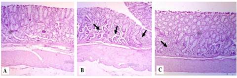
Fig. 1: Representative micrograph showing the comparison of histological damages in colonic epithelium of LG, HG and HG-Se diets in goats. Colon from each group was processed for histological evaluation: colon section of LG diet group (A, scale bar = 100 μm); HG diet group (B, scale bar = 100 μm) and HG-Se diet group (C, scale bar = 100 μm) at 10X magnification. Representative histological sections of the colon tissue were stained by H&E. The arrows indicate damages to colonic epithelial mucosa.
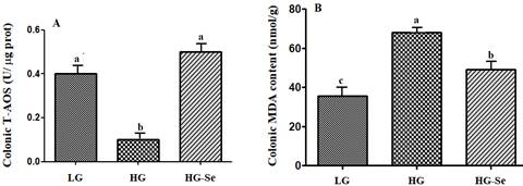
Fig. 2: The effect of LG, HG and HG-Se diets on total antioxidant status (T-AOS: A) and melondialdehyde (MDA: B) concentration in colonic epithelium of goat
The values are mean ± S.E and a, b, c different letters on the bars exhibit the differences between groups with P < 0.05
As in the previous report (Samo et al. 2020) our results showed increased fermentation rate with immediate decrease in pH in the colon of goats, resulting in increased LPS concentration in colon and plasma of the goat fed high grain and high grain-Se diets as compared to low grain diet in current study. The diet induced decrease in luminal pH results in the lysis of gram-negative bacteria that ultimately enhance LPS concentration in the GIT. Acidic medium and LPS level alters the integrity of GIT wall hence, LPS translocation takes place through permeable membrane (Abaker et al. 2017). The diet rich in grain at the rate of 60-90% causes increase in colonic and blood LPS concentration in the goat (Wang et al. 2021a, b). Furthermore, Liu et al. (2013) found that in male goat fed 65% grain diet signifies the outflow of LPS with 0.860.20 Eu/mL LPS concentration in the blood. In present study though the level of LPS increased in both high grain and high grain-Se diet, yet the addition of Se in high grain diet attenuated LPS concentration in colon and plasma of the goats. Shah et al. (2022) in her study have reported similar findings in the goat fed grain rich diet supplemented with Se. Supplementation of Se attenuates the production of LPS and amplifies epithelial integrity in the colon of the goat under diet-induced stress (Samo et al. 2020) also the heat stressed pig jejunum (He et al. 2022). It is suggested that Se supplementation reduces LPS concentration by lowering the mechanism of bacteriolysis, hence lowered the accumulation of luminal free LPS and translocation from GIT. It is suggested that when supplemented with grain rich diet, organic Se in ruminant microflora is incorporated and retained much higher to inorganic Se (Mainville et al. 2009), hence increase the antioxidant stability of microbes that eventually influence them to survive in harmful acidic environment (Čobanová et al. 2017).
Colon histopathology and oxidative stress
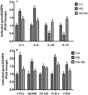
Fig. 3: The effect of LG, HG and HG-Se diets on inflammatory signaling genes in colonic epithelium of goat
The gene expression of interleukins (A) and cytokines (B) were calculated with real time PCR in comparison with GAPDH. Abbreviations: Interluekins (IL), tumer necrotic factor (TNFα), myeloid differentiation factor (MyD88), nuclear factor kappa B (NF-ķB), toll like receptors (TLR-4) and cluster of differentiation (CD14). The values are mean ± S.E and a, b, c different letters on the bars exhibit the differences between groups with P < 0.05
The goats fed high grain diet are at high risk of damages to gastrointestinal integrity (Liu et al. 2013), epithelium of colon being single layered is easily compromised by abnormal luminal environment than multilayered epithelium of the rumen (Malhi et al. 2013). The existing study represents that ten weeks continuous feeding high grain diet to the young goats exhibited severe damages to the mucosal tissue of colon characterized crypt necrosis and inflammatory cell infiltration as compared to the goats fed low grain diet. Luminal acidity and LPS concentration are the factors determining the status of epithelial barrier and the severity of the injury in the tissue of rumen and colon (Emmanuel et al. 2007). Number of studies have reported that decrease in pH due to high grain diet causes epithelial disruption in cecum and colon resulting in outflow of LPS to the blood (Klevenhusen et al. 2013; Tao et al. 2015). In contrast, extra Se supplementation with high grain diet exerted cyto-protective effect by minimizing the colonic epithelial damages. Ameliorative effect of Se have been reported against hepatic cellular injury induced by carbon tetrachloride in rats (Bitiren et al. 2010), aortic injury induced by lipid rich diet in rabbits (Mehta et al. 2002), histo-architectural changes in spleen (Wang et al. 2013), thymus (Chen et al. 2013) and bursa of fibricous (BF) (Chen et al. 2014) induced by aflatoxin (AFB1) in broilers (Hu et al. 2018). It is demonstrated that pH 5.5-7.0 results in cellular apoptosis (Lan et al. 2007). Our previous study confirmed that the goats fed high grain and high grain-Se had relative pH values of 5.8 and 6.1, respectively in colonic digesta (Samo et al. 2020). Since Se supplementation attenuated LPS concentration in the colonic lumen and hence translocation, therefore, in present study minor injury to the epithelium of High grain-Se diet might be due to lowered pH in the colon.
Under normal physiological condition reactive oxygen and nitrogen species (ROS and RNS) such as O2-, H2O2, ROO·, NO, and ONOO- are generated (Yu et al. 2015). However, high level of ROS/RNS adversely alter DNA and oxidizes lipids and proteins in several tissues resulting in cellular damages (Nita and Grzybowski 2016). On the other hand, living beings defend themselves against reactive species via antioxidant defense mechanism that implies the balance between production and elimination of ROS/RNS. This defense system comprises glutathione peroxidase (GSH-Px), superoxide dismutase (SOD) and catalase (CAT) enzymes. The inconsistency between ROS generation and antioxidant defense system marks an OS. Simultaneously, increase in reactive species causes overproduction of melondialdehyde (MDA) as a result of lipid peroxidation thus used as an important marker of OS (Teama 2018). The present findings confirm the presence of OS in goats fed the high grain diet, as indicated by an increased MDA concentration and a reduction in T-AOS levels in the colon. The results presented over here shows compatibility with Abaker et al. (2017) in the liver and Guo et al. (2017) in the plasma of the cows fed high grain diet. Similar to our outcomes, previous studies have investigated that acidic pH and unnecessary production of LPS develops OS by elevated MDA and decreased antioxidant status in gut epithelial tissue thereby aggravates the injury. On the contrary, Se supplemented high grain diet showed increased T-AOS and decreased MDA concentration in high grain-Se as compared to high grain diet. It is well-established fact that additional supplementation with Se reduces concentration of LPS, hence mitigates colonic damage in goats due to OS (Samo et al. 2020) by supporting antioxidation process in blood of heat stressed sheep and pigs (Chen et al. 2020; Mousaie 2021). Se supplementation might have improved Se status in the tissue that ultimately caused alleviation of High grain diet induced OS and epithelial injury in colon.
Plasma acute phase response and inflammation in colon
Lipopolysacchride (LPS) is suggested to be a potent stimulator of proinflammatory response. LPS translocated into the blood results in peripheral blood LPS concentration and engenders reactive oxygen, nitrogen intermediates and bioactive lipids, which indices systemic acute phase response (Khafipour et al. 2009). The initial stage of LPS binds to LPS binding protein (LBP) called 60 kDa APP to initiate inflammation. Additionally, Hp and SAA are well-recognized APPs in the blood that are upregulated in response to various stress conditions, including trauma, infection, and grain/concentrate-rich diets. Therefore they are collectively used as markers of inflammation in ruminant (Alsemgeest et al. 1994). It is described that high grain challenge stimulates acute phase response (APR) signifying by elevation of LBP, SAA and Hp in dairy cows accompanied by the transfer of LPS from the digestive tracts into plasma (Khafipour et al. 2009). In present study, associated with elevated plasma LPS concentration, high grain diet provokes the activation of blood LBP, SAA and Hp in goats. In liver of goats, sheep, and cattle affected by LPS, the gene expression of APPs is altered (Wang et al. 2017; Shah et al. 2022). In contrast, extra Se supplementation mitigated high grain diet induced APPs in the plasma of goat. Consistently, higher levels of Se supplementation beyond the recommended threshold (> 0.5 mg kg⁻¹ diet) help to enhance immune response by the APPs regulation thus reduce postpartum stress (Gong and Xiao 2021) also, mitigate metabolic disorders associated with heat stress in pigs (Liu et al. 2021).
Inflammation is a protective response against harmful stimuli and conditions such as infection of microorganisms and tissue injury plays a vital role in process of host defense and tissue repair. However, uncontrolled inflammatory responses results in excessive or long-term tissue damages supporting the development of acute or chronic inflammatory conditions. The combination/complex of LPS-LBP binds to cluster of differentiation (CD14) which triggers toll like receptor- 4 (TLR-4) and activates innate immunity. The MyD88-dependent signaling pathway, triggered downstream of TLR-4 activation, can lead to the activation of nuclear factor kappa B (NF-κB), which in turn promotes the production of cytokines and other pro-inflammatory mediators (Ryu et al. 2019). Our findings demonstrated that the activation of CD14, TLR-4, MYD88 and NF-κB validates the initiation of inflammatory response in the colon of High grain diet fed goats, in contrast to the goats fed low grain diet. In good agreement with our findings, Tao et al. (2015) in the colon of goats and Guo et al. (2017) in the liver of dairy cows showed similar results through gene expression. Upon stimulation, NF-κB stimulates pro-inflammatory Tumor necrotic factor (TNF-α), interleukin 1 (IL-1) and IL-6 (Bryant et al. 2017) and causes pathophysiological series of events parallel to endotoxaemia or septic shock. Additionally, derange values of APPs in ruminants fed concentrate diets are suggested to be concomitant with alteration in the level of inflammatory cytokines in blood (Chang et al. 2015; Ohtaki et al. 2020). Earlier studies in the liver of goats and cattle revealed that grain rich diet-generated LPS upregulates the NF-κB mRNA expressions, alters the expression of TNF-α, IL-1, IL-6, and IL-10, and subsequently stimulates the production of APPs (Duanmu et al. 2016; Guo et al. 2017). In the present study, goats fed a high-grain diet showed elevated levels of pro-inflammatory cytokines TNF-α, IL-1, and IL-6, along with decreased levels of anti-inflammatory cytokines IL-10 and IL-13 in the colon. These findings suggest that LPS derived from the high grain diet may activate the TLR-4–MyD88 signaling pathway through NF-κB (Shah et al. 2022).
On the contrary, supplemented extra-Se subsided colonic inflammatory response by attenuation of CD14, TLR-4 and MYD88 accompanied with decreased TNF-α, IL-1 and IL-6 values. Simultaneously, increased values of anti-inflammatory IL-10 and IL-13 were observed in colonic epithelium of goats as compared to grain rich diet without Se supplementation in current study. Similar results have been shown in liver of goats fed extra Se with high grain diet studied by Shah et al. (2022). Furthermore, a study in jejunum and thymus of growing pigs subjected to heat and drug-induced stress, suggested that Se supplementation promotes an anti-inflammatory by playing role in inhibition of TLR-4 pathway (He et al. 2022; Liu et al. 2021) as well as in the liver and uterus of mice and chickens challenged with LPS (Al-Dossari et al. 2020; Chen et al. 2020; Qu et al. 2020). Additionally, IL-10 and IL-13 are identified as key intracellular regulators of inflammation, playing a protective role against hepatic injury caused by ischemia in mice (Kato et al. 2003). The specific mechanism behind anti-inflammatory role of Se against LPS-mediated endometritis in rats is due to its role in stimulation of LxRα-ABCA1 pathway followed by cholesterol degradation, which interferes in lipid raft formation and led to the inhibition of TLR-4 migration to lipid rafts (Chen et al. 2020).
Conclusion
Our findings validated that high grain diet increased LPS levels in the colon and blood, triggering local and systemic inflammation leading to epithelial disruption in the colon. The inflammation in the colon was accompanied with TLR-4- MYD88 signaling pathway. This research extends further to comprehend the role of Se as nutrient as well as an anti- stressor in modulating the injurious effects of high grain diet produced-LPS in colon. Moreover, even at higher doses, Se showed beneficial effects without harming animal health under normal or stressed conditions. Se mitigated high grain diet-induced stress and inflammation, reducing colonic injury in goats. This study highlights dual role of Se as a nutrient and anti-stressor in countering LPS-related damage from high grain diets.
Acknowledgments
The first author acknowledges the facilitation provided by Sindh Agriculture University Tandojam i.e., experimental station, laboratory work and technical support.
Author Contributions
MCM and JS planned research work. MAS and FAU interpreted results. NBS analyzed results statistically while NBS and SPS made write up and illustration.
Conflict of Interest
All the authors declared no conflict of interest.
Data Availability
Data presented in this study will be available on a fair request to the all authors.
Ethical Approval
Not applicable to this paper, however the animals were slaughtered according to Islamic law by Halal method.
References
Abaker J, T Xu, D Jin, G Chang, K Zhang, X Shen (2017). Lipopolysaccharide derived from the digestive tract provokes oxidative stress in the liver of dairy cows fed a high-grain diet. J Dairy Sci 100:666‒678
Liu L, D Chen, B Yu, Y Luo, Z Huang, P Zheng, X Mao, J Yu, J Luo, H Yan (2021). Influences of selenium-enriched yeast on growth performance, immune function, and antioxidant capacity in weaned pigs exposure to oxidative stress. BioMed Res Intl 2021:1-11
Razo-Rodriguez O, J Ramirez-Bribiesca, R Lopez-Arellano, A Revilla-Vazquez, S Gonzalez-Munoz, M Cobos-Peralta, L Hernandez-Calva, L McDowell (2013). Effects of dietary level of selenium and grain on digestive metabolism in lambs. Czech J Anim Sci 58:253‒261
Ryu YS, KA Kang, MJ Piao, MJ Ahn, JM Yi, YM Hyun, SH Kim, MK Ko, CO Park, JW Hyun (2019). Particulate matter induces inflammatory cytokine production via activation of NFκB by TLR5-NOX4-ROS signaling in human skin keratinocyte and mouse skin. Redox Biol 21:101080
Stranges S, AG Tabak, E Guallar, MP Rayman, TN Akbaraly, M Laclaustra, G Alfthan, H Mussalo-Rauhamaa, JS Viikari, OT Raitakari (2011). Selenium status and blood lipids: the cardiovascular risk in Young Finns study. J Intern Med 270:469‒477
Tao S, P Tian, Y Luo, J Tian, C Hua, Y Geng, R Cong, Y Ni, R Zhao (2017). Microbiome-metabolome responses to a high-grain diet associated with the hind-gut health of goats. Front Microbiol 8:1-13
Tao S, J Tian, R Cong, L Sun, Y Duanmu, H Dong, Y Ni, R Zhao (2015). Activation of cellular apoptosis in the caecal epithelium is associated with increased oxidative reactions in lactating goats after feeding a high-concentrate diet. Exp Physiol 100:278‒287
Tao S, Y Duanmu, H Dong, Y Ni, J Chen, X Shen, R Zhao (2014). High concentrate diet induced mucosal injuries by enhancing epithelial apoptosis and inflammatory response in the hindgut of goats. PLoS One 9:1-9
Online : 1814-9596
Print : 1560-8530