


Indexing


Journal Cover

Identification and Pathogenicity of Ganoderma Causing Basal Stem Rot in Oil Palm in South Sumatra
Abstract
Basal stem rot is a significant disease affecting oil palm (Elaeis guineensis Jacq.) in Indonesia, with occurrences reported in the Sumatra and Kalimantan regions. Understanding the pathogenic characteristics of fungal pathogens is essential for accurate diagnosis and effective disease management. This research was conducted with an aim to characterize the pathogenicity of three Ganoderma isolates and to identify them using molecular methods. The results showed that the KM8 isolate exhibited the highest pathogenicity, with a disease incidence of 73%, compared to 67% for B55 and 47% for B93. The mycelium of KM8 also demonstrated the fastest growth rate, averaging 0.5 cm day-1, followed by B93 at 0.4 cm day-1 and B55 at 0.15 cm day-1. Symptoms of basal stem rot began to appear three days after inoculation. Cross-sectional observations of the stem revealed brown discoloration in infected plants. Microscopic examination showed clear differences between healthy and Ganoderma-infected tissues, with infected plantlets displaying vascular shrinkage and abnormalities. The isolates were morphologically pre-identified as belonging to genus Ganoderma based on distinctive colony traits and pigmentation. Therefore, species specific primers targeting G. boninense (90F-443R and 90F-444R6) were used to confirm identity at the species level. PCR amplification produced a band of approximately 370 base pairs. All the three isolates were identified as Ganoderma boninense, with a sequence similarity of approximately 98–99%.
Keywords: Disease incidence; Fungal pathogen; Ganoderma boninense; Molecular identification; Plantlet
Identification and Pathogenicity of Ganoderma Causing Basal Stem Rot in Oil Palm in South Sumatra
Pratiwi Ayu Hardini1,2†, Adi Bayu Prakoso3, Safira Medina3, Gregorius Baskara1, Ruli Wandri1†*, Dwi Asmono1, Suwandi Suwandi2, Siti Subandiyah3 and A Muslim2
1Research and Development Department, PT Binasawit Makmur, Sampoerna Agro, Jl. Basuki Rahmat, 7888, Palembang, South Sumatra 30127, Indonesia
2Plant Science Graduate Program, Faculty of Agriculture, Univesitas Sriwijaya, Jl. Padang Selasa 524, Palembang, South Sumatera 310139, Indonesia
3Department of Plant Pest and Disease, Faculty of Agriculture, Gadjah Mada University, Jl. Flora, Bulaksumur, Yogyakarta 55281, Indonesia
*For correspondence: ruli.wandri@sampoernaagro.com
†Contributed equally to this work and are co-first authors
Received 03 July 2025; Accepted 09 August 2025; Published online 22 September 2025
Editor: Arshad Javaid
Abstract
Basal stem rot is a significant disease affecting oil palm (Elaeis guineensis Jacq.) in Indonesia, with occurrences reported in the Sumatra and Kalimantan regions. Understanding the pathogenic characteristics of fungal pathogens is essential for accurate diagnosis and effective disease management. This research was conducted with an aim to characterize the pathogenicity of three Ganoderma isolates and to identify them using molecular methods. The results showed that the KM8 isolate exhibited the highest pathogenicity, with a disease incidence of 73%, compared to 67% for B55 and 47% for B93. The mycelium of KM8 also demonstrated the fastest growth rate, averaging 0.5 cm day-1, followed by B93 at 0.4 cm day-1 and B55 at 0.15 cm day-1. Symptoms of basal stem rot began to appear three days after inoculation. Cross-sectional observations of the stem revealed brown discoloration in infected plants. Microscopic examination showed clear differences between healthy and Ganoderma-infected tissues, with infected plantlets displaying vascular shrinkage and abnormalities. The isolates were morphologically pre-identified as belonging to genus Ganoderma based on distinctive colony traits and pigmentation. Therefore, species specific primers targeting G. boninense (90F-443R and 90F-444R6) were used to confirm identity at the species level. PCR amplification produced a band of approximately 370 base pairs. All the three isolates were identified as Ganoderma boninense, with a sequence similarity of approximately 98–99%.
Keywords: Disease incidence; Fungal pathogen; Ganoderma boninense; Molecular identification; Plantlet
Introduction
Basal stem rot represents one of the most serious diseases affecting oil palm in Indonesia (Paterson 2023). The country's tropical climate, driven by its geographical location, provides favorable conditions for the development of this disease (Jazuli et al. 2022). The causative agent is a fungus belonging to the genus Ganoderma (Zakaria 2023). Basal stem rot can reduce income by up to USD 350 per year and decrease fresh fruit bunch yields by 50–80% (Paterson and Lima 2019). Early screening of oil palm genetic materials for Ganoderma tolerance is increasingly critical due to the persistent spread of basal stem rot in major production areas. In South Sumatera, the disease has become a major constraint for replanting program, as infection are often undetected during early stages and may persist in the soil or planting material (Paterson 2019). Current plantations face difficultly in identifying symptomless but infected individuals, which contribute to the re-emergence of the disease even after sanitation efforts (Khoo and Chong 2023).
Species of the genus Ganoderma are commonly found and pose major problems in oil palm plantations in Indonesia (Hamzah et al. 2022). Ganoderma includes many species that are distributed across oil palm-growing regions. Some well-known pathogens that cause basal stem rot include Ganoderma boninense, G. zonatum, and G. miniatocinctum (Bharudin et al. 2022). In regions such as North Sumatra, South Sumatra, and Kalimantan, the incidence of basal stem rot caused by G. boninense has been reported (Wijayanti et al. 2024; Saputra et al. 2025). Morphologically, this fungus is very similar to other Ganoderma species, which are saprophytic fungi. Field surveys often result in misdiagnosis due to the morphological similarity of the fruiting bodies, or basidiocarps (Khoo and Chong 2023). Therefore, to support the accurate implementation of control measures, molecular identification is necessary (Adotey et al. 2023).
In general, Ganoderma is known as a subtle pathogen, however, under certain environmental conditions such as monoculture planting, extreme weather changes, and imbalances in soil microbiota, it can become a severe threat, exhibiting aggressive growth (Karunarathna et al. 2024). Although Ganoderma boninense is recognized as the main causal of basal stem rot in oil palm, intra-species variation in aggressiveness among isolates from different regions remain understudied. Environmental factors, host-pathogen interactions, and genetic variability can result in differential virulence among isolates (Khoo and Chong 2023; Lekete-Lawson et al. 2025). Therefore, characterizing the pathogenicity of each isolate is essential to identify the most aggressive strain, which can be prioritized for further resistance screening and control strategy development. Due to the long-life cycle of oil palm, symptom observation for basal stem rot may require a considerable amount of time. Thus, conventional pathogenicity testing using field seedlings requires extended observation periods (up to 6 months or more) and is often affected by uncontrolled environmental factors. In contrast, oil palm plantlets derived from somatic embryogenesis offer a rapid, uniform, and contamination-free model system that allows early detection of disease symptoms. Moreover, plantlets with lignin content are more susceptible to fungal invasion, making them a suitable model for differentiating isolate virulence (Rencoret et al. 2021; USDA 2024). Therefore, this study aimed to compare the pathogenicity of three Ganoderma isolates from South Sumatra, and to identify these isolates using molecular techniques to confirm their taxonomy.
Materials and Methods
Isolation of Ganoderma
Three isolates (KM 8, B55 and B93) of Ganoderma used in this study were isolated from basidiocarps on basal stem rot-affected oil palm at different plantations in Ogan Komering Ilir, South Sumatera, Indonesia. Freshly collected basidiocarps were surfaced sterilized, cut and plated on PDA medium. Mycelium growing from the plated tissue were purified to be pure culture and maintain on PDA.
Identification of Ganoderma isolate
The isolates were then characterized based on their morphological appearance and mycelial growth on PDA. The color of the aerial mycelium, surface texture, color changes in the basal plate, and colony margin were observed to assess morphological characteristics (Xing et al. 2008). Mycelial growth was measured daily by recording the colony diameter. Colony growth rate was calculated by dividing the colony diameter by the number of days until the PDA plate was fully covered.
The pure culture of Ganoderma was then subjected to molecular identification to confirm that the isolate was G. boninense. DNA was extracted from the pure culture using the Zymo Research Quick-DNA™ Fungal/Bacterial Miniprep Kit. After obtaining the DNA template, amplification was carried out using a thermal cycler with G. boninense-specific primers, 90F-443R and 90F-444R6 (Hardini et al. 2023). The PCR amplification consisted of 25 cycles: initial denaturation at 96°C for 5 min; denaturation at 94°C for 3 min; annealing at 60°C for 1.5 min. and extension, followed by a final extension at 72°C for 5 min. The amplified PCR products were then sent to 1st BASE Genomics (Singapore) for sequencing. The resulting sequences were compared to reference strains of G. boninense in the NCBI database (https://www.ncbi.nlm.nih.gov/).
Pathogenicity test
The identified G. boninense isolates were refreshed on Potato Dextrose Agar (PDA) and incubated at room temperature for 14 days until full mycelial coverage was observed on the Petri dish. The pathogenicity test was conducted using oil palm plantlets. The plantlets were prepared at the Tissue Culture Laboratory of PT Sampoerna Agro Tbk, using susceptible material for Ganoderma (Dura genotype). They were cultivated on Murashige and Skoog (MS) medium without activated charcoal to facilitate root observation during Ganoderma infection. The plantlets used for the test met the following criteria: more than three months old, uniform height, more than three leaves, and free from contamination.
The isolates were then inoculated into the oil palm plantlets for pathogenicity testing, to ensure that the symptoms resulting from artificial infection matched typical basal stem rot symptoms. Inoculation was performed under aseptic conditions inside a biosafety cabinet. Plantlets were grouped based on the treatment, including three isolate codes (KM8, B55 and B93) and a non-inoculated control. Each isolate was applied to 15 plantlets as replicates. Inoculation was conducted by placing three 0.5-mm diameter PDA plugs containing actively growing G. boninense around the base of each stem. The culture tubes were then sealed, and incubation was carried out at 27°C. Observations continued for up to 40 days post-infection. Disease symptoms were assessed using a scoring method described by Susanto et al. (2013) as shown in Table 1.
Table 1: Scoring criteria for symptoms assessing
|
Scoring |
Symptom Description |
|
0 |
Healthy plant/asymptomatic |
|
1 |
Necrotic on leaves ≤ 25% |
|
2 |
Necrotic on leaves ≤ 50% |
|
3 |
Necrotic on 2-3 leaves and mycelium colonizing the roots, necrotic ≤ 50% |
|
4 |
The plant becomes necrotized and dies, mycelium colonize roots, stems and leaves |
Table 2: Result of BLAST analysis on specific amplicon sequencing results
|
Sample |
Description |
Identity (%) |
Accession number |
|
B93_90F_443R |
Ganoderma boninense isolate MD 12 A |
99.40 |
MW647647 |
|
B55_90F_443R |
Ganoderma boninense isolate MD 12 A |
99.10 |
MW647639 |
|
KM8_90F_443R |
Ganoderma boninense isolate MD 12 A |
98.40 |
OQ435807.1 |
The scoring data is then calculated for disease incidence (DI) and disease severity (Masnilah et al. 2020):

Disease severity (DS) was calculated per individual plant in the treatment with the following formula severity (Masnilah et al. 2020):
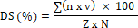
Where, n : Total of infested plants with a certain category
v : Scale value of each attack category
N : Total of plants observed
Z : Highest scale value.
All statistical analyses were performed using SPSS version 25.0. Data on disease incidence and disease severity were analyzed using one-way analysis of variance (ANOVA), followed by Duncan’s Multiple Range Test (DMRT) to identify significant differences among Ganoderma isolates at the 5% probability level. Results were considered statistically significant at P < 0.05.
Observations were also made on a cross-section of the stem of infected oil palms to determine the condition of the inside of the stem. The incision starts from the base of the stem to the end of the stem near the lowermost leaf axil. Furthermore, the damaged tissue was observed with a light microscope with 400 magnifications (Rees et al. 2009) After obtaining data on isolates that are suspected of having the highest level of pathogenicity with symptoms as a basal stem rot.
Results
Isolation and Identification of Ganoderma
Colony morphology of three Ganoderma isolates viz., KM8, B55 and B93, grown on potato dextrose agar (PDA) for 14 days is shown in Fig. 1, all isolates exhibited distinguishable colony appearances in terms of color, texture, and pigmentations. Isolate B55 formed a compact white colony with fine hyphal strands on the surface. Pigmentation developed progressively in the center, resulting in a dark brown coloration by day 14. Isolate B39 showed a more diffuse white colony with radial hyphal growth. A central brownish zone was visible in the later stage of incubation. Isolate KM8 exhibited a dense white mycelial mat visible yellowish pigmentation distributed across the surface. The appearance of the colonies different among the isolates, particularly in color intensity and colony margin structure.
Molecular identification
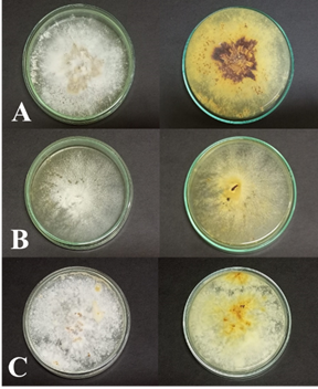
Fig. 1: Morphological appearance of three Ganoderma isolates grown on potato dextrose agar (PDA): (A) B55, (B) B93 and (C) KM8
PCR results using both primer pairs, 90F-443R and 90F-444R6, successfully amplified the G. boninense target gene specifically, producing amplicons of approximately 370 bp (Fig. 2). The results of sequencing analysis presented in Table 2 show that isolates B55, B93 and KM8 are G. boninense species with a percentage identity value of ~99%.
Growth rate of Ganoderma isolates
KM8 exhibited the highest growth rate (0.5 cm day-1), followed by B93 (0.4 cm day-1), and B55 (0.15 cm day-1), demonstrated the slowest growth rate. The differences became more apparent from day 6 to 10. All isolates showed minimal radial growth during the initial 3 days. Growth began to increase progressively from day 4 onward. The differences in colony diameter became more pronounced starting from day 7, with KM8 reaching the highest recorded diameter by day 14. Isolates B55 and B93 showed growth rates throughout the observation period (Fig. 3).
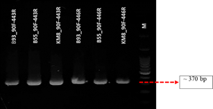
Fig. 2: Electrophoresis results of PCR amplicons using specific primers 90F-443R and 90F-446R
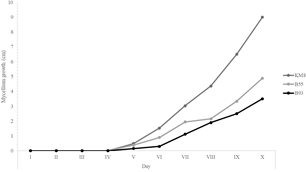
Fig. 3: Radial mycelial growth (cm) of three Ganoderma isolates on PDA
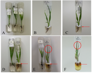
Fig. 4: Ganoderma boninense infection on oil palm plantlets; (A) 0 DAI, (B) 3 DAI, (C) 7 DAI, (D) 10 DAI, (E) 17 DAI and (F) 38 DAI
G. boninense infection in oil palm plantlets
Infection of G. boninense isolates in in vitro oil palm plantlets showed that all three isolates simultaneously induced symptoms of basal stem rot (BSR) beginning at 3 days after inoculation (DAI). The disease incidence and severity were as follows: plantlets infected with isolate KM8 exhibited the highest disease incidence at 73% and a severity of 76%; isolate B55 showed a disease incidence of 67% and a severity of 70%; while isolate B93 had the lowest incidence at 47% and severity at 46% (Fig. 4).
Initial symptoms appeared at 3 DAI, characterized by hyphal colonization at the stem base, early signs of stem rot, and leaf necrosis covering less than 25% of the leaf area. By 7 DAI, symptoms progressed with leaf necrosis exceeding 50%, and more extensive decay observed at the roots and stem base. At 10 DAI, leaf desiccation was evident, along with discoloration of the growth medium and visible fungal hyphae on leaf surfaces. By the third week, mycelial growth became abundant, and stem and root decay became increasingly severe. At 38 DAI, all plantlets had died (Fig. 5).
Microscopic examination of transverse sections stained with lactophenol cotton blue demonstrated intact and well-structured vascular networks in non-infected plants. In contrast, G. boninense-infected tissues (Fig. 6) displayed damaged and collapsed vascular vessels, indicating disruption of the plant’s transport system caused by fungal invasion.
Disease incidence and disease severity of Ganoderma
Table 3 presents disease incidence and disease severity recorded from three Ganoderma isolates (KM8, B55, dan B 93) after inoculation on oil palm plantlets. In terms of disease incidence, isolate KM8 resulted in the highest percentage (73%), followed by B55 (64%) and B93 (46%). Disease severity also followed a similar trend, with KM8 producing the highest score (76%) and B93 showing the lowest (45%). The control group didn’t exhibit any symptoms of disease.
Discussion
The comprehensive identification and pathogenicity evaluation of Ganoderma isolated from basal stem rot (BSR)-affected oil palm in South Sumatra provides critical insights for improving early detection and disease management strategies. Morphological assessments underlined phenotypic diversity among isolates, which manifested as variations in colony pigmentation, mycelial density, and structural margins. However, morphological identification alone is inadequate due to the high phenotypic plasticity of Ganoderma under varying environmental conditions (Purnamasari et al. 2012).
Table 3: Disease incidence and disease severity of Ganoderma in oil palm plantlets
|
Isolate |
Disease Incidence (%) |
Disease Severity (%) |
|
KM8 |
73 a |
76 a |
|
B55 |
64 b |
70 b |
|
B93 |
46 c |
45 c |
|
K |
0 d |
0 d |
|
Mean |
60.89 |
63.67 |
*values followed by different letters in each column are significantly different at P < 0.05
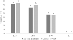
Fig. 5: Disease incidence and disease severity in plantlets at 38 day after inoculation. Bars with the same letter are not significantly different according to Duncan’s Multiple Range Test (DMRT) at P ≤ 0.05
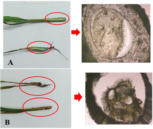
Fig. 6: Microscopic observation of (A) non-infected treatment plantlets showing healthy tissue; (B) Ganoderma infected treatment plantlets and infected tissue (200x magnification)
These morphological differences in colony appearance—including variations in mycelial density, color and pigment distribution—reflect phenotypic diversity among the isolates and may be indicative of differences in physiological activity and virulence (Khoo and Chong 2023). Therefore, the integration of molecular techniques—particularly PCR using species-specific primers (90F-443R and 90F-444R6)—provided robust confirmation that all three isolates belong to Ganoderma boninense, with high sequence similarity (>98%) to GenBank references (Adotey et al. 2023; Hardini et al. 2023).
The use of plantlets derived from somatic embryogenesis as an infection model enabled rapid and controlled assessment of isolate aggressiveness, circumventing the limitations associated with field seedlings such as long growth cycles and environmental variability. This model was particularly effective given that in vitro-grown plantlets typically exhibit lower lignin content, thereby rendering them more vulnerable to fungal colonization (Rencoret et al. 2021). Additionally, the absence of activated charcoal in the MS medium facilitated clearer observation of root colonization and disease progression (Visnovsky et al. 2020). In contrast, the use of older seedlings or field-grown oil palm present several limitations when assessing infection. First, the latency period for symptom development in mature seedlings is considerably longer, often requiring 8 to 12 weeks or more (Idris 2004). Second, the presence of well- developed lignifoed tissues in older seedlings enhances their structural resistance, potentially masking early fungal colonization and leading to underestimation of severity in less aggressive isolates.
In this study, the Dura genotype was selected due to its well-documented susceptibility to G. boninense and its wide usage in oil palm breeding programs (Purba et al. 2012; Swaray et al. 2020). The high disease incidence observed in this genotype is consistent with previous reports associating inbreeding depression in DxD crosses with increased vulnerability to Ganoderma infection (Corley and Tinker 2003). Anatomical responses such as increased root proliferation, tissue necrosis, and vascular collapse observed in infected plantlets reinforce the physiological stress response mechanisms, possibly including structural adaptations to biotic stress (Akmalia and Pranatami 2021).
Comparative analysis of disease incidence and severity clearly indicated that pathogenicity varies significantly among isolates, confirming intra-species virulence diversity as suggested by Karunarathna et al. (2024). These findings validate the need to evaluate individual isolate aggressiveness when developing resistant plant materials. Furthermore, the early emergence of necrotic symptoms and vascular degradation highlights the efficiency of the plantlet-based assay for preliminary resistance screening and pathogen characterization, particularly when rapid results are required.
Histological observation corroborated earlier work by Rees et al. (2009) and Senewe et al. (2023), who demonstrated that Ganoderma infection leads to shrinkage and disruption of vascular tissues, impeding water and nutrient flow and ultimately causing plant death. The discoloration of culture media observed during infection could reflect secondary metabolite secretion by the pathogen or plant-derived stress responses—both of which are important hallmarks of host–pathogen interaction (Muslim 2019).
The use of artificial inoculation under aseptic conditions ensured experimental reproducibility and reliability. This methodological consistency is vital for future screening of oil palm varieties with moderate tolerance to G. boninense, offering a scalable and resource-efficient alternative to conventional field trials (Susanto and Prasetyo 2013).
This study reaffirms the importance of integrating morphological, molecular, and pathogenicity assessments for accurate and functional characterization of Ganoderma boninense. The observed differences in virulence among isolates emphasize the necessity of isolate-specific analysis when developing mitigation strategies, especially in high-risk replanting zones in Indonesia’s major oil palm-producing regions (Paterson 2023; Saputra et al. 2025).
Conclusion
Ganoderma isolates obtained from PT Binasawit Makmur collection were confirmed as the causal agents of basal stem rot in oil palm, as evidenced by the development of characteristic symptoms in pathogenicity assays. Among the tested isolates, KM8 exhibited the highest virulence, indicated by more rapid and severe symptom progression compared to other isolates. Molecular identification using ITS region sequencing revealed that all three isolates belonged to Ganoderma boninense with sequences similarities exceeding 98%.
Acknowledgements
The authors gratefully acknowledge PT Bina Sawit Makmur (Sampoerna Agro) for providing research materials and technical facilities. We also thank the Faculty of Agriculture, Universitas Sriwijaya, and the Department of Plant Pests and Diseases, Gadjah Mada University, for their support in molecular identification. The contributions of all individuals who assisted in this research are sincerely appreciated.
Author Contribution
PAH, GB, RW and DA: Designed the study, providing plantlets materials and Ganoderma isolates, conducted research and wrote the research paper. ABP, SM and SS: Provided laboratory facility, build up methods, helped analysis and writing the manuscript while SW and AM: Reviewed paper and data analysis.
Conflict of Interest
All authors declare no conflict of interest.
Data Availability
Data presented in this study will be available on a fair request to corresponding author.
Ethic Approval
Not applicable to this paper.
References
Adotey G, RN Alolga, A Quarco, P Yerenkyi, P Out, AK Anang, LKN Okine, J Gbewonyo, JC Holliday, VC Lombardi (2023). Molecular identification and characterization of five Ganoderma species from the lower volta river basin of Ghana based on nuclear ribosomal DNA (nrDNA) sequences. J Fung 10:1-12
Akmalia HA, DA Pranatami (2021). Biologi Umum Untuk Mahasiswa. Alinea Media Dipantara
Bharudin I, AFFA Wahab, MAA Samad, NX Yie, MA Zairun, FD Abu Bakar, AMA Murad (2022). Review update on the life cycle, plant-microbe interaction, genomics, detection and control strategies of the oil palm pathogen Ganoderma boninense. Biology 11:1-18
Corley RHV, PB Tinker (2003). The Oil Palm, 4th edn. Blackwell Publishing, Oxford, UK
Hamzah A, R Saputra, F Puspita, B Nasrul, Irfandri, NS Depari (2022). Ganoderma diversity from smallholder oil plantation in peatlands of Kampar district, Indonesia based on mycelia morphology and somatic incompatibility. Biodiversitas 22:16–22
Hardini PA, IB Nugroho, AB Prakoso, GBA Nugraha, A Wibowo, A Widiastuti, WD Sawitri, A Soffan, R Wandri, S Subandiyah, D Asmono (2023). DNA barcoding of Ganoderma boninense Pat. Isolated from Oil Plantation in South Sumatera, Indonesia. International Palm Oil Congress (PIPOC), MPOB-Malaysia
Idris AS (2004). The identity of Ganoderma species responsible for BSR and upper stem rot in oil palm plantation of Malaysia. Malaysia Palm Oil Board (MPOB)
Jazuli NA, K Kamu, KP Chong, D Gabda, A Hassan, IA Seman, CM Ho (2022). A review of factors affecting Ganoderma basal stem rot disease progress in oil palm. Plants 11:1-14 doi:10.3390/plants11192462
Karunarathna SC, NM Patabendige, W Lu, S Asad, KK Hapuarachchi (2024). An in-depth study of phytopathogenic Ganoderma: pathogenicity, advanced detection techniques, control strategies, and sustainable management. J Fung 10:1-78
Khoo YW, KP Chong (2023). Ganoderma boninense: general characteristics of pathogenicity and methods of control. Front Plant Sci 14:1-17
Lekete-Lawson E, GCVD Pujie, EA Osekre, FK Ackah (2025). First report of Ganoderma ryvardenii causing basal stem rot (BSR) disease on oil palm (Elaeis guineensis Jacq.) in Ghana. F100Research 14:1-26
Masnilah R, WS Wahyuni, S Dwi, A Majid (2020). Insidensi dan keparahan penyakit penting tanaman padi di Kabupaten Jember. Agritop J Ilm Pertan 18:1–12
Muslim A (2019). Pengendalian Hayati Patogen Tanaman Dengan Mikroorganisme Antagonis. UNSRI Press
Paterson RRM (2019). Ganoderma boninense disease of oil palm to significantly reduce production after 2050 in Sumatera if projected climate change occurs. Microorganism 7:1-8
Paterson RRM (2023). Future climate effect on basal stem rot of conventional and modified oil palm in Indonesia and Thailand. Forests 14:1-13
Paterson RRM, N Lima (2019). Ecology and biotechnology of thermophilic fungi on crops under global warming. In: Fungi Extreme Environmental Ecology Role Biotechnology, pp:81–96. Springer
Purba AR, U Setiawati, A Susanto, M Rahmaningsih, Y Yenni, HY Rahmadi, SPC Nelson (2012). Indonesia’s Experience of Developing Ganoderma-Tolerant/Resistant Oil Palm Planting Material. ISOPB, Cartagena, Colombia
Purnamasari MI, C Prihatna, AW Gunawan, A Suwanto (2012). Isolation and molecular identification of Ganoderma spp. associated with basal stem rot disease in oil palm. J Fitopatol Indo 8:9-15
Rees RW, J Flood, Y Hasan, U Potter, RM Cooper (2009). Basal stem rot of oil palm (E. guineensis): Mode of root infection and lower stem invasion by G. boninense. Plant Pathol 8:982–989
Rencoret J, A Gutierrez, G Marques, JCD Rio, Y Tobimatsu, PY Lam, MP Boada, FJR Duenas, JM Barassa, AT Martinez (2021). New insight on structure forming the lignin-like fractions of ancestral plants. Front Plant Sci 12:1-14
Saputra MA, B Yuniasih, EN Kristalisasi (2025). Intensitas serangan penyakit busuk pangkal batang pada perkebunan kelapa sawit. Agroforetech 3:27–33
Senewe RE, M Pasireron, B Sahetapy (2023). Penyakit busuk pangkal batang tanaman kelapa sawit oleh patogen Ganoderma spp. J Trop Agric 1:65–73
Susanto A, AE Prasetyo (2013). Metode skrinning tanaman kelapa sawit toleran Ganoderma. Warta PPKS 18:105–114
Susanto A, AE Prasetyo, H Priwiratama, S Wening (2013). Ganoderma boninense penyebab penyakit busuk pangkal batang atas kelapa sawit. J Fitopatol Indo 9:123-126
Swaray S, MD Amirudin, MY Rafii, S Jamian, MF Ismail, M Jalloh, M Marjuni, MM Mohammad, O Yusuff (2020). Influence of parental dura and pisifera genetic origins on oil palm fruit set ratio and yield components in their D x P progenies. Agronomy 10:1-30
USDA (2024). Plant Pathogen Confirmatory Diagnostic Laboratory. Animal and Plant Health Inspection Service, US Department of Agriculture
Visnovsky P, P Panda, KR Everett, A Lu, RC Butler, RK Taylor, AR Pitman (2020). A PCR diagnostic assay for rapid detection of plant pathogenic pseudomonads. Plant Pathol 69:1311–1330
Wijayanti E, AE Prasetyo, H Priwiratama, TAP Rozziansha, DD Eris, AS Mulyatni, AF Lubis, SP Rambe (2024). Kejadian penyakit busuk pangkal batang pada tanaman kelapa sawit menjelang tanam ulang di Sumatera Utara bagian barat. Warta PPKS 29:45–60
Xing JH, YF Sun, YL Han, BK Cui, YC Dai (2008). Morphological and molecular identification of two new Ganoderma species on Casuarina equisetifolia from China. MycoKeys 34:93–108
Zakaria L (2023). Basal Stem Rot of Oil Palm: The Pathogen, Disease Incidence, and Control Methods. APS Online Publications. https://doi.org/10.1094/PDIS-02-22-0358-FE
Online : 1814-9596
Print : 1560-8530